Explore
 Validate
 Learn
Learn Western blot
Antibody data
- Antibody Data
- Antigen structure
- References [4]
- Comments [0]
- Validations
- Western blot [3]
- Immunocytochemistry [3]
- Immunohistochemistry [3]
- Other assay [3]
Submit
Validation data
Reference
Comment
Report error
- Product number
- PA5-79479 - Provider product page

- Provider
- Invitrogen Antibodies
- Product name
- IL-18 Polyclonal Antibody
- Antibody type
- Polyclonal
- Antigen
- Recombinant full-length protein
- Description
- Reconstitute with 0.2 mL of distilled water to yield a concentration of 500 µg/mL. Positive Control - WB: human Hela whole cell, human HepG2 whole cell. IHC: human tonsil tissue. ICC/IF: A549 cell.
- Reactivity
- Human
- Host
- Rabbit
- Isotype
- IgG
- Vial size
- 100 μg
- Concentration
- 500 μg/mL
- Storage
- -20°C
Submitted references Tongxinluo May Alleviate Inflammation and Improve the Stability of Atherosclerotic Plaques by Changing the Intestinal Flora.
Subversion of GBP-mediated host defense by E3 ligases acquired during Yersinia pestis evolution.
Huaier extract suppresses non-small cell lung cancer progression through activating NLRP3-dependent pyroptosis.
LncRNA MALAT1 promoted high glucose-induced pyroptosis of renal tubular epithelial cell by sponging miR-30c targeting for NLRP3.
Qi Y, Liu W, Yan X, Zhang C, Zhang C, Liu L, Zheng X, Suo M, Ti Y, Ni M, Zhang M, Bu P
Frontiers in pharmacology 2022;13:805266
Frontiers in pharmacology 2022;13:805266
Subversion of GBP-mediated host defense by E3 ligases acquired during Yersinia pestis evolution.
Cao S, Jiao Y, Jiang W, Wu Y, Qin S, Ren Y, You Y, Tan Y, Guo X, Chen H, Zhang Y, Wu G, Wang T, Zhou Y, Song Y, Cui Y, Shao F, Yang R, Du Z
Nature communications 2022 Aug 4;13(1):4526
Nature communications 2022 Aug 4;13(1):4526
Huaier extract suppresses non-small cell lung cancer progression through activating NLRP3-dependent pyroptosis.
Xie J, Zhuan B, Wang H, Wang Y, Wang X, Yuan Q, Yang Z
Anatomical record (Hoboken, N.J. : 2007) 2021 Feb;304(2):291-301
Anatomical record (Hoboken, N.J. : 2007) 2021 Feb;304(2):291-301
LncRNA MALAT1 promoted high glucose-induced pyroptosis of renal tubular epithelial cell by sponging miR-30c targeting for NLRP3.
Liu C, Zhuo H, Ye MY, Huang GX, Fan M, Huang XZ
The Kaohsiung journal of medical sciences 2020 Sep;36(9):682-691
The Kaohsiung journal of medical sciences 2020 Sep;36(9):682-691
No comments: Submit comment
Supportive validation
- Submitted by
- Invitrogen Antibodies (provider)
- Main image

- Experimental details
- Western blot analysis of IL-18 in HeLa whole cell lysate using 40 µg per well. Sample was incubated with IL-18 (Product # PA5-79479) at a dilution of 0.5 µg/mL.
- Submitted by
- Invitrogen Antibodies (provider)
- Main image

- Experimental details
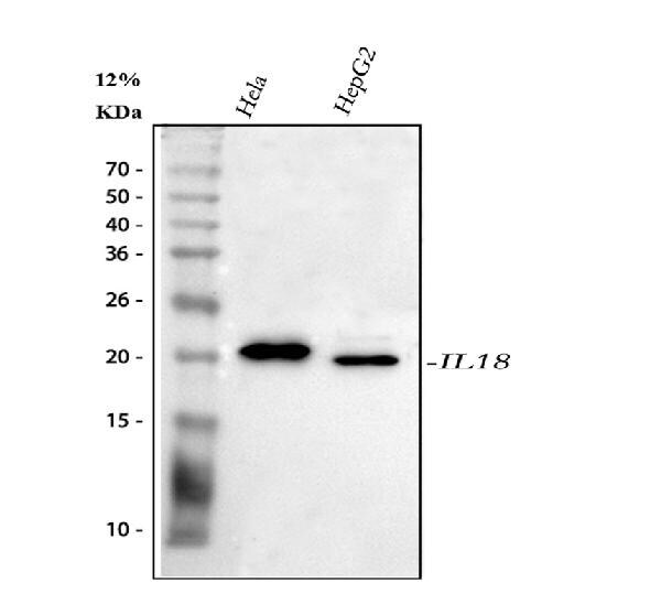
- Western blot analysis of IL-18 using IL-18 Polyclonal Antibody (Product # PA5-79479). Electrophoresis was performed on a 5-20% SDS-PAGE gel at 70V (Stacking gel)/90V (Resolving gel) for 2-3 hours. The sample well of each lane was loaded with 30 µg of sample under reducing conditions. Lane 1: human Hela whole cell lysates. Lane 2: human HepG2 whole cell lysates. After electrophoresis, proteins were transferred to a nitrocellulose membrane at 150 mA for 50-90 minutes. Blocked the membrane with 5% non-fat milk/TBS for 1.5 hour at RT. The membrane was incubated with IL-18 Polyclonal Antibody at 0.5 µg/mL overnight at 4°C, then washed with TBS-0.1%Tween 3 times with 5 minutes each and probed with a goat anti-rabbit IgG-HRP secondary antibody at a dilution of 1:5,000 for 1.5 hour at RT. The signal is developed using an Enhanced Chemiluminescent detection (ECL) kit with Tanon 5200 system. A specific band was detected for IL-18 at approximately 22 kDa. The expected band size for IL-18 is at 22 kDa.
- Submitted by
- Invitrogen Antibodies (provider)
- Main image
- Experimental details
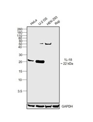
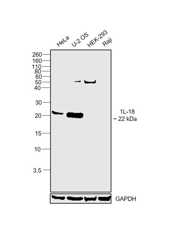
- Western blot was performed using Anti-IL-18 Polyclonal Antibody (Product # PA5-79479) and a 22 kDa band corresponding to Interleukin-18 was observed across cell lines tested except HEK-293 and Raji which is reported to be low. Whole cell extracts (30 µg lysate) of HeLa (Lane 1), U-2 OS (Lane 2), HEK-293 (Lane 3) and Raji (Lane 4) were electrophoresed using NuPAGE™ 12% Bis-Tris Protein Gel (Product # NP0342BOX). Resolved proteins were then transferred onto a Nitrocellulose membrane (Product # IB23001) by iBlot® 2 Dry Blotting System (Product # IB21001). The blot was probed with the primary antibody (0.2 µg/mL) and detected by chemiluminescence with Goat anti-Rabbit IgG (Heavy Chain) Superclonal™ Recombinant Secondary Antibody, HRP (Product # A27036, 1:4000) using the iBright FL 1000 (Product # A32752). Chemiluminescent detection was performed using Novex® ECL Chemiluminescent Substrate Reagent Kit (Product # WP20005).
Supportive validation
- Submitted by
- Invitrogen Antibodies (provider)
- Main image

- Experimental details
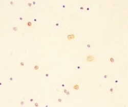
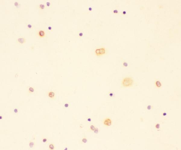
- Immunocytochemistry analysis of IL18 using anti-IL18 antibody(Product # PA5-79479). IL18 was detected in an immunocytochemical section of blood cells. Enzyme antigen retrieval was performed using IHC enzyme antigen retrieval reagent for 15 mins. The cells were blocked with 10% goat serum and then incubated with 1 μg/mL rabbit anti-IL18 antibody (Product # PA5-79479) overnight at 4°C. Biotinylated goat anti-rabbit IgG was used as secondary antibody and incubated for 30 minutes at 37°C. The section was developed using Strepavidin-Biotin-Complex (SABC) with DAB as the chromogen.

- Submitted by
- Invitrogen Antibodies (provider)
- Main image
- Experimental details
- Immunocytochemistry analysis of IL18 using anti-IL18 antibody(Product # PA5-79479). IL18 was detected in an immunocytochemical section of blood cells. Enzyme antigen retrieval was performed using IHC enzyme antigen retrieval reagent for 15 mins. The cells were blocked with 10% goat serum and then incubated with 1 μg/mL rabbit anti-IL18 antibody (Product # PA5-79479) overnight at 4°C. Biotinylated goat anti-rabbit IgG was used as secondary antibody and incubated for 30 minutes at 37°C. The section was developed using Strepavidin-Biotin-Complex (SABC) with DAB as the chromogen.
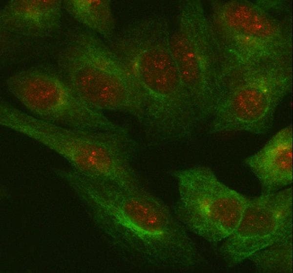
- Submitted by
- Invitrogen Antibodies (provider)
- Main image
- Experimental details
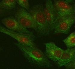
- Immunocytochemistry/Immunofluorescence analysis of IL-18 in A549 cell using IL-18 Polyclonal Antibody (Product # PA5-79479). Enzyme antigen retrieval was performed using IHC enzyme antigen retrieval reagent for 15 mins. The cells were blocked with 10% goat serum and incubated with the primary antibody at 5 µg/mL and mouse anti-Beta Tubulin antibody overnight at 4°C. Cy3 conjugated goat anti-rabbit IgG and DyLight 488 conjugated goat anti-mouse IgG were used as secondary antibody at 1:100 dilution and incubated for 30 minutes at 37°C. Visualized using a fluorescence microscope and filter sets appropriate for the label used.
Supportive validation
- Submitted by
- Invitrogen Antibodies (provider)
- Main image
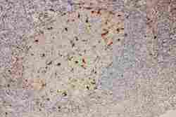
- Experimental details
- Immunohistochemistry analysis of IL-18 on paraffin-embedded human tonsil tissue. Sample was incubated with IL-18 polyclonal antibody (Product# PA5-79479).
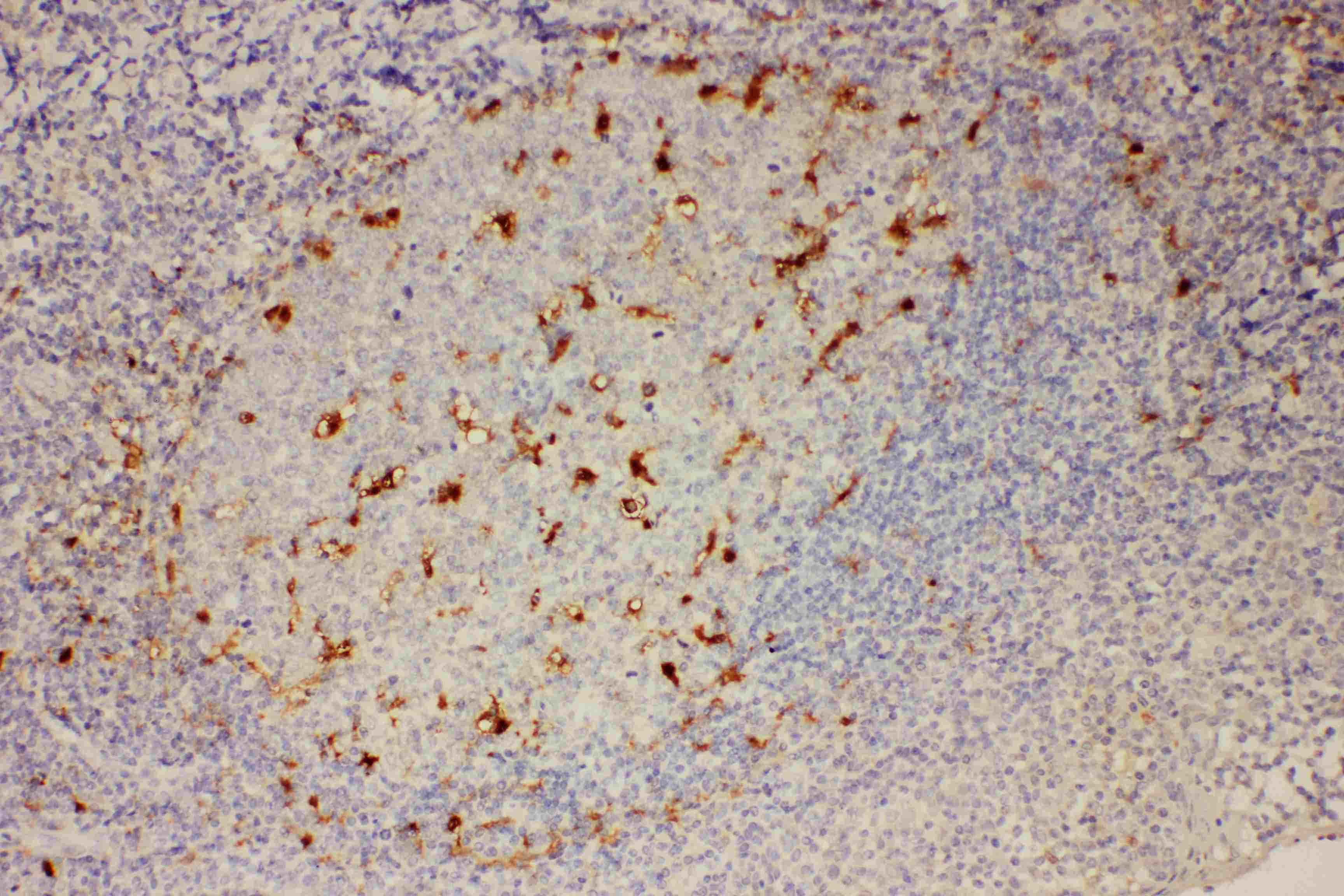
- Submitted by
- Invitrogen Antibodies (provider)
- Main image
- Experimental details
- Immunohistochemistry (Paraffin) analysis of IL-18 in paraffin-embedded section of human tonsil tissue using IL-18 Polyclonal Antibody (Product # PA5-79479). Heat mediated antigen retrieval was performed in EDTA buffer (pH 8.0, epitope retrieval solution). The tissue section was blocked with 10% goat serum. The tissue section was then incubated with the primary antibody at a 2 µg/mL dilution overnight at 4°C. Peroxidase conjugated goat anti-rabbit IgG was used as secondary antibody and incubated for 30 minutes at 37°C. The tissue section was developed using HRP Conjugated Rabbit IgG Super Vision Assay Kit with DAB as the chromogen.
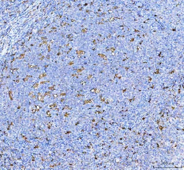
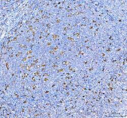
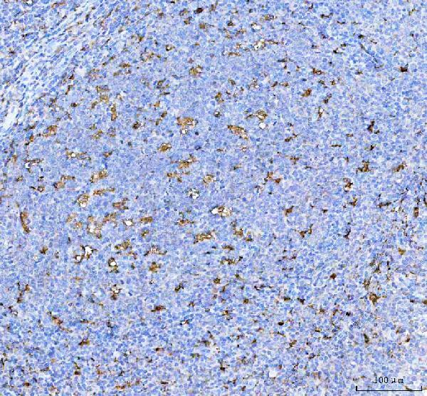
Supportive validation
- Submitted by
- Invitrogen Antibodies (provider)
- Main image
- Experimental details
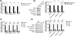
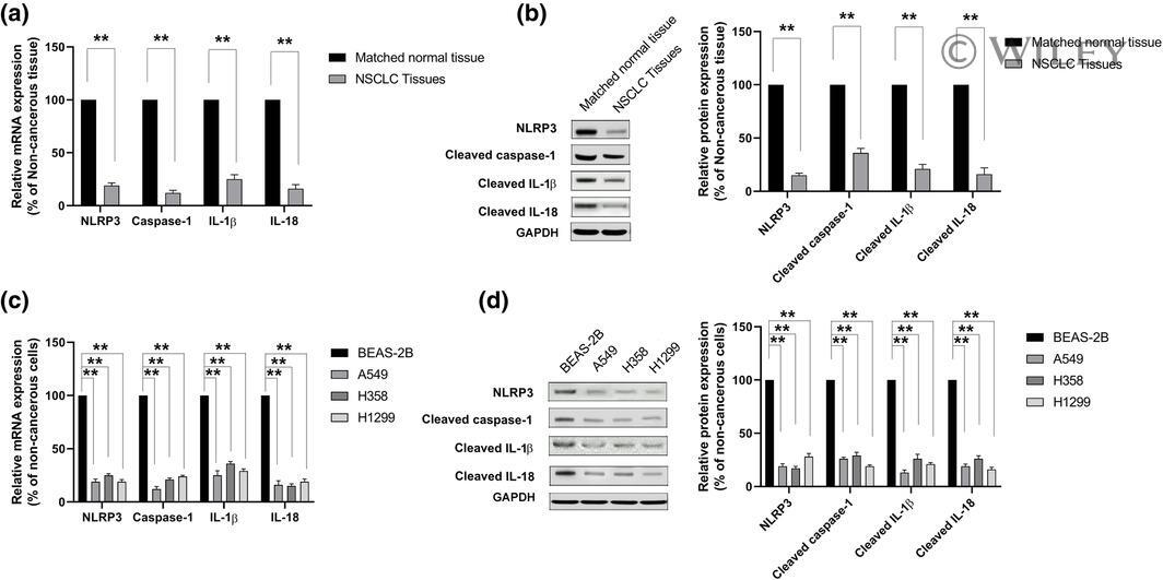
- 1 FIGURE Pyroptosis levels were decreased in non-small cell lung cancer (NSCLC) tissues and cell lines. (a and c) The mRNA levels of pyroptotic-related genes (NLRP3, caspase-1, IL-1beta, and IL-18) in NSCLC tissues, adjacent noncancerous tissues, NSCLC cell lines (A549, H358, and H520), and normal lung epithelial cell line BEAS-2B were analyzed by using qRT-PCR. (b and d) The proteins levels of pyroptotic-related genes (NLRP3, caspase-1, IL-1beta, and IL-18) in NSCLC tissues, adjacent noncancerous tissues, NSCLC cell lines (A549, H358, and H520), and normal lung epithelial cell line BEAS-2B were analyzed by using western blot. GAPDH is chosen as protein controls. ** p < .01
- Submitted by
- Invitrogen Antibodies (provider)
- Main image

- Experimental details
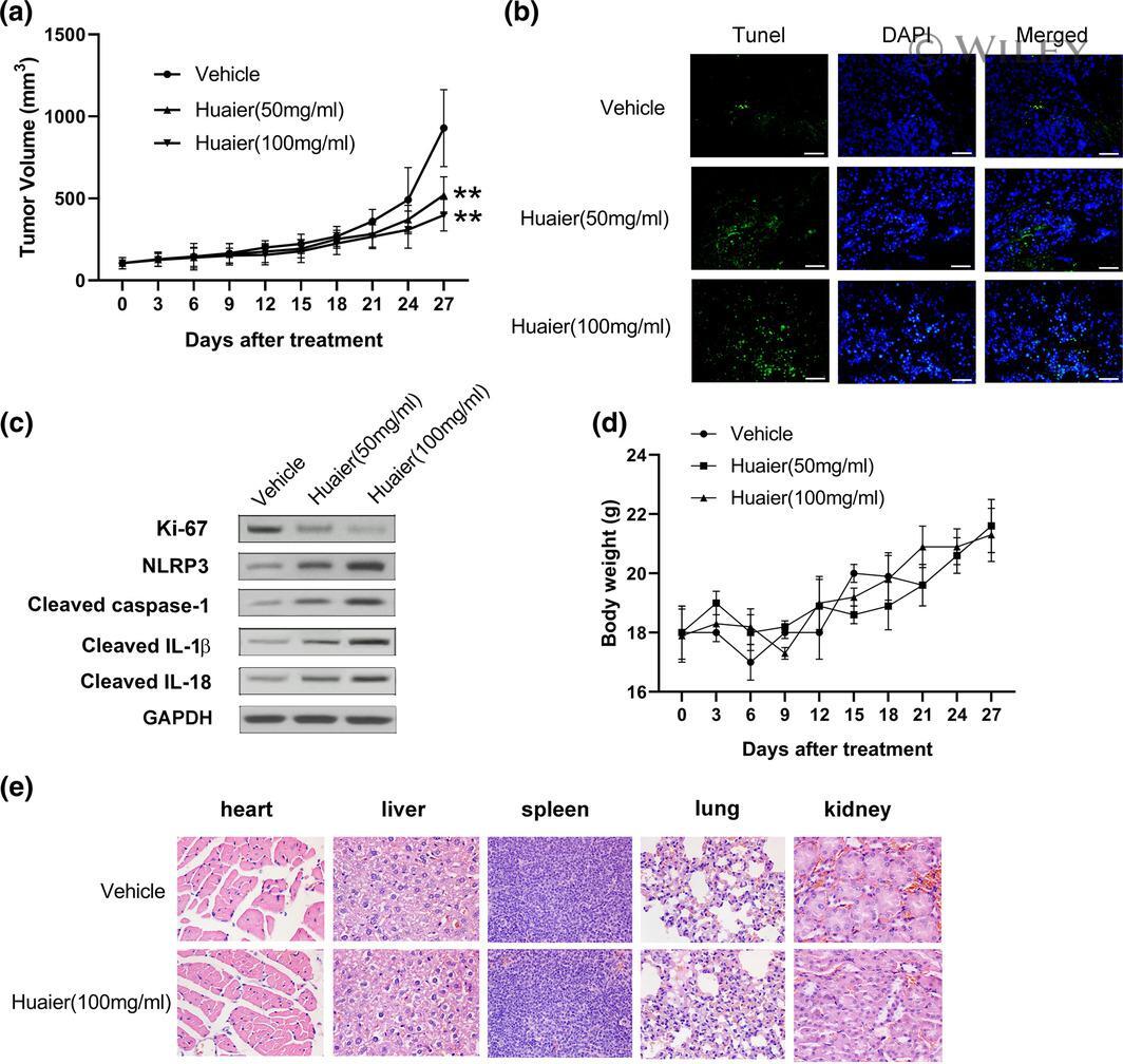
- 6 FIGURE Huaier suppresses tumor growth and induces pyroptosis in vivo. Tumorigenesis assay was performed by subcutaneous injection of H520 cells into flanks of BALB/c nude mice. (a) Tumor volumes were measured at the indicated days post-injection. (b) The effect of huaier on apoptosis in tumor tissues was evaluated by TUNEL assay. (c) The western blot analysis detected the relative expression of Ki-67, NLRP3, caspase-1, IL-1beta, and IL-18 in tumor tissues. (d) Bodyweight of mice in the vehicle group, low dose group (50 mg/ml), and high dose group (100 mg/ml) were evaluated every 3 days. (e) H&E staining was applied to evaluate the changes in the heart, liver, spleen, lung, and kidney tissues. ** p < .01
- Submitted by
- Invitrogen Antibodies (provider)
- Main image

- Experimental details
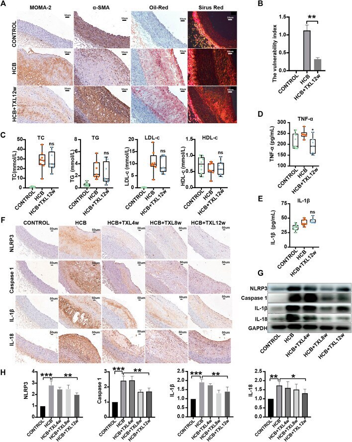
- FIGURE 2 Tongxinluo improved the stability of HCB-induced plaques and alleviated inflammation through non-lipid-dependent pathways in rabbits. CONTROL: normal diet (n = 12); HCB: high cholesterol diet and balloon injury (n = 8); HCB + TXL 4w: HCB and treated with TXL for 4 weeks (n = 7). HCB + TXL 8w: HCB and treated with TXL for 8 weeks (n = 8). HCB + TXL 12w: HCB and treated with TXL for 12 weeks (n = 10) (A) Plaque's stability test results. Oil red O staining was applied to show lipid in abdominal aorta. Sirius red staining was used to detect collagen fibers. Abdominal aorta sections were stained for macrophages (MOMA-2) or vascular smooth muscle cells (alpha-SMA). bar = 50 mum (B) Plaque vulnerability index statistics. Data are shown as the mean +- S.E.M. **: p < 0.01. (C,D,E) The changes of Blood lipids (C), Serum inflammatory factors TNF-alpha (D) andIL-1beta (E) of New Zealand White Rabbit in normal diet (CONTROL, n = 12), high cholesterol diet (HCB, n = 8) and high cholesterol diet with TXL treatment for 12 weeks (HCB + TXL 12w, n = 10), measured by enzymatic determination. Non-parametric test was used for statistical tests. Data were presented as median (minimum to maximum) (F) Histopathological results. Immunohistological staining of the abdominal aortic cross-section showing dense, positive NLRP3/Caspase-1/IL-1beta/IL-18 in the five different treatment groups. (G,H) Western blotting to quantitate NLRP3/Caspase-1/IL-1beta/IL-18 protein expression (G) , and non-param