Explore
 Validate
 Learn
Learn Western blot
Antibody data
- Antibody Data
- Antigen structure
- References [0]
- Comments [0]
- Validations
- Western blot [5]
- Immunocytochemistry [1]
- Immunohistochemistry [5]
Submit
Validation data
Reference
Comment
Report error
- Product number
- RQ5347 - Provider product page

- Provider
- NSJ Bioreagents
- Product name
- HAPLN1 Antibody
- Antibody type
- Monoclonal
- Description
- This highly specific HAPLN1 antibody is suitable for use in Immunohistochemistry/Western blot/Immunofluorescence applications with human and mouse samples.
- Reactivity
- Human, Mouse
- Host
- Rabbit
- Conjugate
- Unconjugated
- Antibody clone number
- BHA-8
- Vial size
- 100 ul
- Concentration
- Antibody in PBS with 0.02% sodium azide, 50% glycerol and 0.4-0.5mg/ml BSA
- Storage
- Store the HAPLN1 antibody at -20oC.
No comments: Submit comment
Supportive validation
- Submitted by
- NSJ Bioreagents (provider)
- Main image

- Experimental details
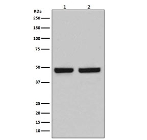
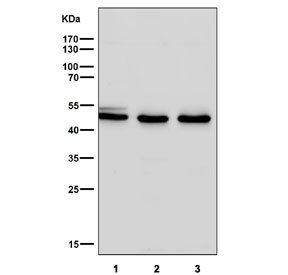
- Western blot testing of 1) human Caco-2 and 2) mouse spleen lysate with HAPLN1 antibody. Expected molecular weight: 41-48 kDa depending on glycosylation level.
- Submitted by
- NSJ Bioreagents (provider)
- Main image
- Experimental details
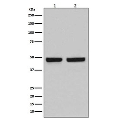
- Western blot testing of 1) human Caco-2 and 2) mouse spleen lysate with HAPLN1 antibody. Expected molecular weight: 41-48 kDa depending on glycosylation level.

- Submitted by
- NSJ Bioreagents (provider)
- Main image
- Experimental details
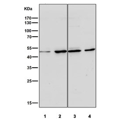
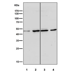
- Western blot testing of human 1) HeLa, 2) Jurkat, 3) MCF7 and 4) SH-SY5Y cell lysate with HAPLN1 antibody at 1:1000. Expected molecular weight: 41-48 kDa depending on glycosylation level.
- Submitted by
- NSJ Bioreagents (provider)
- Main image
- Experimental details
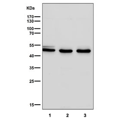
- Western blot testing of human 1) A549, 2) NCI-H1299 and 3) HUVEC lysate with HAPLN1 antibody at 1:1000. Expected molecular weight: 41-48 kDa depending on glycosylation level.
- Submitted by
- NSJ Bioreagents (provider)
- Main image

- Experimental details
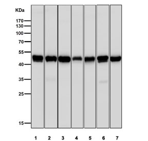
- Western blot testing of 1) mouse liver, 2) mouse spleen, 3) mouse brain, 4) rat heart, 5) rat liver, 6) rat kidney and 7) rat brain tissue lysate with HAPLN1 antibody at 1:1000. Expected molecular weight: 41-48 kDa depending on glycosylation level.
Supportive validation
- Submitted by
- NSJ Bioreagents (provider)
- Main image
- Experimental details
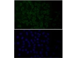
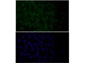
- Immunofluorescent staining of FFPE human HeLa cells with HAPLN1 antibody (green) and DAPI nuclear stain (blue). HIER: steam section in pH6 citrate buffer for 20 min.
Supportive validation
- Submitted by
- NSJ Bioreagents (provider)
- Main image
- Experimental details
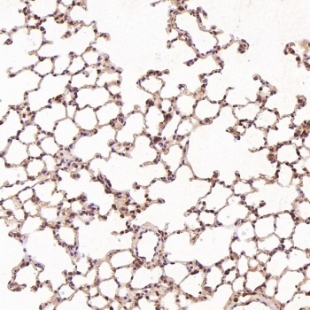
- IHC staining of FFPE human spleen tissue with HAPLN1 antibody. HIER: boil tissue sections in pH6, 10mM citrate buffer, for 10-20 min and allow to cool before testing.
- Submitted by
- NSJ Bioreagents (provider)
- Main image
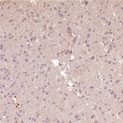
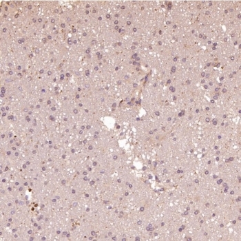
- Experimental details
- IHC staining of FFPE human glioblastoma tissue with HAPLN1 antibody at 1:50. HIER: boil tissue sections in pH6 citrate buffer for 20 min and allow to cool before testing.
- Submitted by
- NSJ Bioreagents (provider)
- Main image
- Experimental details
- IHC staining of FFPE mouse heart tissue with HAPLN1 antibody at 1:50. HIER: boil tissue sections in pH6 citrate buffer for 20 min and allow to cool before testing.
- Submitted by
- NSJ Bioreagents (provider)
- Main image
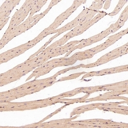
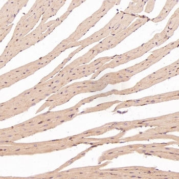
- Experimental details
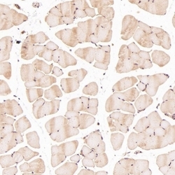
- IHC staining of FFPE mouse skeletal muscle tissue with HAPLN1 antibody at 1:50. HIER: boil tissue sections in pH6 citrate buffer for 20 min and allow to cool before testing.

- Submitted by
- NSJ Bioreagents (provider)
- Main image

- Experimental details
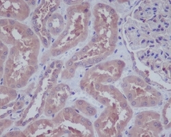
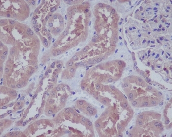
- IHC staining of FFPE rat liver tissue with HAPLN1 antibody at 1:50. HIER: boil tissue sections in pH6 citrate buffer for 20 min and allow to cool before testing.