 Explore
 Validate
 Learn
Learn Western blot
 Immunocytochemistry
Antibody data
- Antibody Data
- Antigen structure
- References [4]
- Comments [0]
- Validations
- Western blot [1]
- Immunocytochemistry [1]
- Immunohistochemistry [1]
Submit
Validation data
Reference
Comment
Report error
- Product number
- AMAb91019 - Provider product page

- Provider
- Atlas Antibodies
- Proper citation
- Atlas Antibodies Cat#AMAb91019, RRID:AB_2665765
- Product name
- Anti-HSP90B1
- Antibody type
- Monoclonal
- Description
- Monoclonal Antibody against Human HSP90B1, Clone ID: CL2647, Gene description: heat shock protein 90kDa beta (Grp94), member 1, Alternative Gene Names: GP96, GRP94, TRA1, Validated applications: WB, IHC, ICC, Uniprot ID: P14625, Storage: Store at +4°C for short term storage. Long time storage is recommended at -20°C.
- Reactivity
- Human
- Host
- Mouse
- Conjugate
- Unconjugated
- Isotype
- IgG
- Antibody clone number
- CL2647
- Vial size
- 100 µl
- Storage
- Store at +4°C for short term storage. Long time storage is recommended at -20°C.
- Handling
- The antibody solution should be gently mixed before use.
Submitted references Tld1 is a regulator of triglyceride lipolysis that demarcates a lipid droplet subpopulation
A comprehensive library of fluorescent constructs of SARS‐CoV‐2 proteins and their initial characterisation in different cell types
Cerebellar ataxia disease–associated Snx14 promotes lipid droplet growth at ER–droplet contacts
Speer N, Braun R, Reynolds E, Brudnicka A, Swanson J, Henne W
Journal of Cell Biology 2024;223(1)
Journal of Cell Biology 2024;223(1)
Paul B, Merta H, Ugrankar-Banerjee R, Hensley M, Tran S, Dias do Vale G, McDonald J, Farber S, Henne W
2024
2024
A comprehensive library of fluorescent constructs of SARS‐CoV‐2 proteins and their initial characterisation in different cell types
Miserey‐Lenkei S, Trajkovic K, D'Ambrosio J, Patel A, Čopič A, Mathur P, Schauer K, Goud B, Albanèse V, Gautier R, Subra M, Kovacs D, Barelli H, Antonny B
Biology of the Cell 2021;113(7):311-328
Biology of the Cell 2021;113(7):311-328
Cerebellar ataxia disease–associated Snx14 promotes lipid droplet growth at ER–droplet contacts
Datta S, Liu Y, Hariri H, Bowerman J, Henne W
Journal of Cell Biology 2019;218(4):1335-1351
Journal of Cell Biology 2019;218(4):1335-1351
No comments: Submit comment
Enhanced validation
- Submitted by
- Atlas Antibodies (provider)
- Enhanced method
- Genetic validation
- Main image

- Experimental details
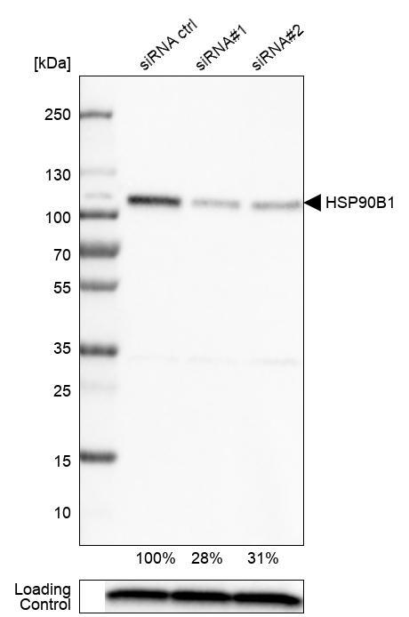
- Western blot analysis in U-251MG cells transfected with control siRNA, target specific siRNA probe #1 and #2, using Anti-HSP90B1 antibody. Remaining relative intensity is presented. Loading control: Anti-PPIB.
- Sample type
- Human
- Protocol
- Protocol
Supportive validation
- Submitted by
- Atlas Antibodies (provider)
- Main image

- Experimental details
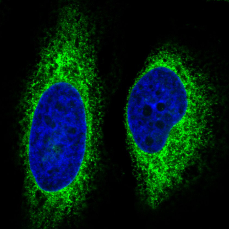
- Immunofluorescence staining in HeLa cell line with Anti-HSP90B1 monoclonal antibody, showing specific staining of endoplasmic reticulum in green. Microtubule- and nuclear probes are visualized in red and blue respectively (where available).
- Sample type
- Human
Supportive validation
- Submitted by
- Atlas Antibodies (provider)
- Enhanced method
- Orthogonal validation
- Main image
- Experimental details
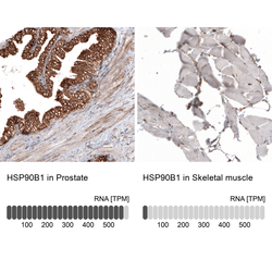
- Immunohistochemistry analysis in human prostate and skeletal muscle tissues using AMAb91019 antibody. Corresponding HSP90B1 RNA-seq data are presented for the same tissues.
- Sample type
- Human
- Protocol
- Protocol
