 Explore
 Validate
 Learn
Learn Western blot
 Immunocytochemistry
Antibody data
- Antibody Data
- Antigen structure
- References [0]
- Comments [0]
- Validations
- Western blot [1]
- Immunoprecipitation [3]
- Immunohistochemistry [1]
Submit
Validation data
Reference
Comment
Report error
- Product number
- LS-C98044 - Provider product page

- Provider
- LSBio
- Proper citation
- LifeSpan Cat#LS-C98044, RRID:AB_2089924
- Product name
- DCK / Deoxycytidine kinase Antibody (aa171-200) LS-C98044
- Antibody type
- Polyclonal
- Description
- Ammonium sulfate precipitation
- Reactivity
- Human, Mouse
- Host
- Rabbit
- Storage
- Maintain refrigerated at 2°C to 8°C for up to 6 months. For long term storage store at -20°C.
No comments: Submit comment
Supportive validation
- Submitted by
- LSBio (provider)
- Enhanced method
- Genetic validation
- Main image

- Experimental details
- The anti-DCK antibody is used in Western blot to detect DCK in mouse intestine tissue lysate.

Supportive validation
- Submitted by
- LSBio (provider)
- Enhanced method
- Genetic validation
- Main image

- Experimental details
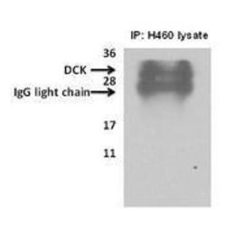
- Deoxycytidine kinase(DCK) immunoprecipitated from H460cells with 7.5ug (microgram) of the dCK antibody using the Pierce classic mammalian IP kit (#45217) reagent as described as manufacturer instructions (lane 1, 3) and Current Protocols in Cell Biology, 1998, 7.2.1-7.2.21. Proteins separated on a 12% SDS gel, transferred to a PVDF membrane and probed with 1:700 dilution of DCK antibody. Bands were detected using enhanced chemiluminescence (SuperSignal West Pico Chemiluminescent Substrate Kit). No specific reagents were employed to remove IgG from immunoprecipitated sample. Data courtesy of Dr. Stacy Shord, University of Illinois, Chicago.

- Submitted by
- LSBio (provider)
- Main image
- Experimental details
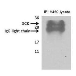
- Deoxycytidine kinase(DCK) immunoprecipitated from H460cells with 7.5ug (microgram) of the dCK antibody using the Pierce classic mammalian IP kit (#45217) reagent as described as manufacturer instructions (lane 1, 3) and Current Protocols in Cell Biology, 1998, 7.2.1-7.2.21. Proteins separated on a 12% SDS gel, transferred to a PVDF membrane and probed with 1:700 dilution of DCK antibody. Bands were detected using enhanced chemiluminescence (SuperSignal West Pico Chemiluminescent Substrate Kit). No specific reagents were employed to remove IgG from immunoprecipitated sample. Data courtesy of Dr. Stacy Shord, University of Illinois, Chicago.

- Submitted by
- LSBio (provider)
- Main image
- Experimental details
- Deoxycytidine kinase(DCK) immunoprecipitated from H460cells with 7.5ug (microgram) of the dCK antibody using the Pierce classic mammalian IP kit (#45217) reagent as described as manufacturer instructions (lane 1, 3) and Current Protocols in Cell Biology, 1998, 7.2.1-7.2.21. Proteins separated on a 12% SDS gel, transferred to a PVDF membrane and probed with 1:700 dilution of DCK antibody. Bands were detected using enhanced chemiluminescence (SuperSignal West Pico Chemiluminescent Substrate Kit). No specific reagents were employed to remove IgG from immunoprecipitated sample. Data courtesy of Dr. Stacy Shord, University of Illinois, Chicago.

Supportive validation
- Submitted by
- LSBio (provider)
- Enhanced method
- Genetic validation
- Main image
- Experimental details
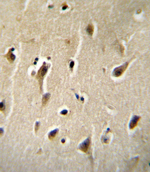
- DCK Antibody immunohistochemistry of formalin-fixed and paraffin-embedded human brain tissue followed by peroxidase-conjugated secondary antibody and DAB staining.
