Explore
 Validate
 Learn
Learn Western blot
 Immunocytochemistry
Antibody data
- Antibody Data
- Antigen structure
- References [1]
- Comments [0]
- Validations
- Western blot [7]
- Immunocytochemistry [2]
- Immunoprecipitation [1]
- Immunohistochemistry [1]
Submit
Validation data
Reference
Comment
Report error
- Product number
- GTX102800 - Provider product page

- Provider
- GeneTex
- Proper citation
- GeneTex Cat#GTX102800, RRID:AB_11163443
- Product name
- DCK antibody
- Antibody type
- Polyclonal
- Reactivity
- Human, Mouse, Rat
- Host
- Rabbit
Submitted references Using genome-wide CRISPR library screening with library resistant DCK to find new sources of Ara-C drug resistance in AML.
Kurata M, Rathe SK, Bailey NJ, Aumann NK, Jones JM, Veldhuijzen GW, Moriarity BS, Largaespada DA
Scientific reports 2016 Nov 3;6:36199
Scientific reports 2016 Nov 3;6:36199
No comments: Submit comment
Enhanced validation
Supportive validation
- Submitted by
- GeneTex (provider)
- Enhanced method
- Genetic validation
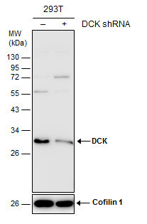
- Main image
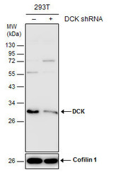
- Experimental details
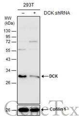
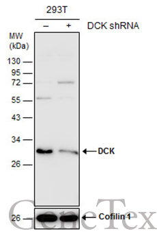
- Non-transfected (¡V) and transfected (+) 293T whole cell extracts (30 ?g) were separated by 12% SDS-PAGE, and the membrane was blotted with DCK antibody (GTX102800) diluted at 1:10000. The HRP-conjugated anti-rabbit IgG antibody (GTX213110-01) was used to detect the primary antibody.
Supportive validation
- Submitted by
- GeneTex (provider)
- Main image
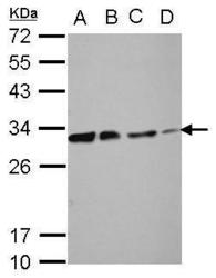
- Experimental details
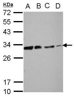
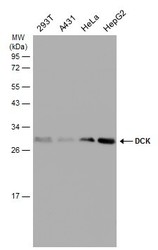
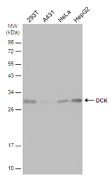
- Sample (30 ?g of whole cell lysate) A: 293T B: A431 C: HeLa D: HepG2 12% SDS PAGE GTX102800 diluted at 1:10000 The HRP-conjugated anti-rabbit IgG antibody (GTX213110-01) was used to detect the primary antibody.
- Submitted by
- GeneTex (provider)
- Main image
- Experimental details
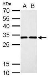
- DCK antibody detects DCK protein by western blot analysis.A. 30 ?g PC-12 whole cell lysate/extract B. 30 ?g Rat2 whole cell lysate/extract 12% SDS-PAGEDCK antibody (GTX102800) dilution: 1:1000 The HRP-conjugated anti-rabbit IgG antibody (GTX213110-01) was used to detect the primary antibody.
- Submitted by
- GeneTex (provider)
- Main image
- Experimental details
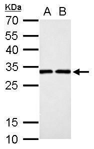
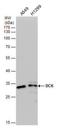
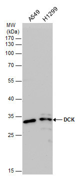
- DCK antibody detects DCK protein by western blot analysis. Various whole cell extracts (30 ?g) were separated by 12% SDS-PAGE, and the membrane was blotted with DCK antibody (GTX102800) diluted at a dilution of 1:5000. The HRP-conjugated anti-rabbit IgG antibody (GTX213110-01) was used to detect the primary antibody.
- Submitted by
- GeneTex (provider)
- Main image
- Experimental details
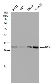
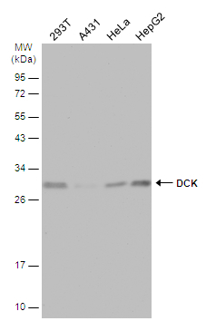
- Various whole cell extracts (30 ?g) were separated by 12% SDS-PAGE, and the membrane was blotted with DCK antibody (GTX102800) diluted at 1:5000. The HRP-conjugated anti-rabbit IgG antibody (GTX213110-01) was used to detect the primary antibody.
- Submitted by
- GeneTex (provider)
- Main image
- Experimental details
- Non-transfected (¡V) and transfected (+) 293T whole cell extracts (30 ?g) were separated by 12% SDS-PAGE, and the membrane was blotted with DCK antibody (GTX102800) diluted at 1:10000. The HRP-conjugated anti-rabbit IgG antibody (GTX213110-01) was used to detect the primary antibody.
- Submitted by
- GeneTex (provider)
- Main image
- Experimental details
- Various whole cell extracts (30 ?g) were separated by 12% SDS-PAGE, and the membrane was blotted with DCK antibody (GTX102800) diluted at 1:5000. The HRP-conjugated anti-rabbit IgG antibody (GTX213110-01) was used to detect the primary antibody.
Supportive validation
- Submitted by
- GeneTex (provider)
- Main image

- Experimental details
- DCK antibody detects DCK protein at nucleus by immunofluorescent analysis.Sample: HeLa cells were fixed in 4% paraformaldehyde at RT for 15 min.Green: DCK protein stained by DCK antibody (GTX102800) diluted at 1:500.Red: Phalloidin, a cytoskeleton marker, diluted at 1:100.Scale bar = 10 £gm.
- Submitted by
- GeneTex (provider)
- Main image

- Experimental details
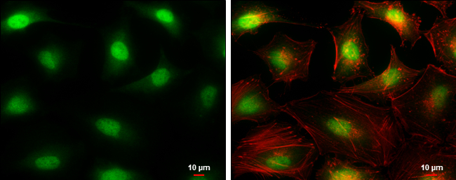
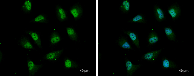
- DCK antibody detects DCK protein at nucleus by immunofluorescent analysis.Sample: HeLa cells were fixed in 4% paraformaldehyde at RT for 15 min.Green: DCK protein stained by DCK antibody (GTX102800) diluted at 1:200.Blue: Hoechst 33342 staining.Scale bar = 10 £gm.
Supportive validation
- Submitted by
- GeneTex (provider)
- Main image
- Experimental details
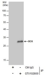
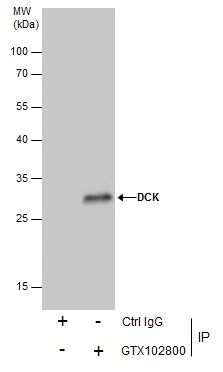
- Immunoprecipitation of DCK protein from 293T whole cell extracts using 5 £gg of DCK antibody (GTX102800).Western blot analysis was performed using DCK antibody (GTX102800).EasyBlot anti-Rabbit IgG (GTX221666-01) was used as a secondary reagent.
Supportive validation
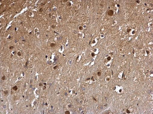
- Submitted by
- GeneTex (provider)
- Main image
- Experimental details
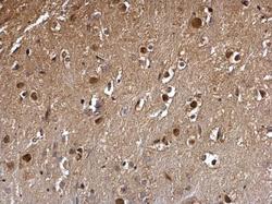
- DCK antibody detects DCK protein at nucleus on mouse hind brain by immunohistochemical analysis. Sample: Paraffin-embedded mouse hind brain. DCK antibody (GTX102800) dilution: 1:500.