Explore
 Validate
 Learn
Learn Western blot
 ELISA
ELISAAntibody data
- Antibody Data
- Antigen structure
- References [19]
- Comments [0]
- Validations
- Western blot [1]
Submit
Validation data
Reference
Comment
Report error
- Product number
- 22287-1-AP - Provider product page

- Provider
- Proteintech Group
- Product name
- MYH3 antibody
- Antibody type
- Polyclonal
- Description
- MYH3 antibody (Cat. #22287-1-AP) is a rabbit polyclonal antibody that shows reactivity with human, mouse and has been validated for the following applications: IP, WB,ELISA.
- Reactivity
- Human, Mouse
- Host
- Rabbit
- Conjugate
- Unconjugated
- Isotype
- IgG
- Vial size
- 20ul, 150ul
Submitted references Bioengineered human pre-vascularized microtissues reconstruct vascular niche and regenerative microenvironment to enhance functional skeletal muscle regeneration.
Activin A secretion by muscle-repairing macrophages induces heterotopic ossification in mice.
LCN2-ACOD1 Signalling Affects the Post-Injury Regeneration of Skeletal Muscle Through Mediating Ferroptosis.
Mechanism of Myh3 gene regulation of intramuscular fat content in Beijing black pigs via the MAPK signaling pathway.
Aminoguanidine hemisulfate improves mitochondrial autophagy, oxidative stress, and muscle force in Duchenne muscular dystrophy via the AKT/FOXO1 pathway in mdx mice.
Intramuscular adipose tissue restricts functional muscle recovery.
Molecular cloning of TPM3 gene in qinchuan cattle and its effect on myoblast proliferation and differentiation.
Myogenic exosome miR-140-5p modulates skeletal muscle regeneration and injury repair by regulating muscle satellite cells.
Hedgehog signaling via its ligand DHH acts as cell fate determinant during skeletal muscle regeneration.
MiR-18 inhibitor promotes the differentiation of bovine skeletal muscle-derived satellite cells by increasing MEF2D expression.
A non-canonical tricarboxylic acid cycle underlies cellular identity.
Alkaline nucleoplasm facilitates contractile gene expression in the mammalian heart.
Distinct MUNC lncRNA structural domains regulate transcription of different promyogenic factors.
Deletion of phosphatidylserine flippase β-subunit Tmem30a in satellite cells leads to delayed skeletal muscle regeneration.
Molecular and Metabolic Mechanism of Low-Intensity Pulsed Ultrasound Improving Muscle Atrophy in Hindlimb Unloading Rats.
miR-206 family is important for mitochondrial and muscle function, but not essential for myogenesis in vitro.
Podocan Promotes Differentiation of Bovine Skeletal Muscle Satellite Cells by Regulating the Wnt4-β-Catenin Signaling Pathway.
TCEA3 promotes differentiation of C2C12 cells via an Annexin A1-mediated transforming growth factor-β signaling pathway.
B4GALNT2 (GALGT2) Gene Therapy Reduces Skeletal Muscle Pathology in the FKRP P448L Mouse Model of Limb Girdle Muscular Dystrophy 2I.
Liang C, Gao Y, Li J, Liang Q, Li M, Yang J, Su X, Zhang J, Tian W, Liao L
Biomaterials 2026 Mar;326:123645
Biomaterials 2026 Mar;326:123645
Activin A secretion by muscle-repairing macrophages induces heterotopic ossification in mice.
Yin W, Okamoto K, Terashima A, Pluemsakunthai W, Ono T, Ito-Kureha T, Akira S, Hashizume Y, Baron R, Ueha S, Matsushima K, Matzuk MM, Mishina Y, Takayanagi H
The Journal of clinical investigation 2026 Mar 2;136(5)
The Journal of clinical investigation 2026 Mar 2;136(5)
LCN2-ACOD1 Signalling Affects the Post-Injury Regeneration of Skeletal Muscle Through Mediating Ferroptosis.
Hao X, Shi H, Wu D, Liang R, Zhao T, Sun W, Wang Y, Yu X, Luo X, Yan Y, Lu J, Wang H, Wang J
Cell proliferation 2026 Apr;59(4):e70130
Cell proliferation 2026 Apr;59(4):e70130
Mechanism of Myh3 gene regulation of intramuscular fat content in Beijing black pigs via the MAPK signaling pathway.
Luo Y, Xu G, Zhang C, Wen Z, Chu H, Dai Y, Hong L, Zhang J, Vignon AS, Sun C, Zhang L, Pu L
BMC genomics 2025 Jul 7;26(1):642
BMC genomics 2025 Jul 7;26(1):642
Aminoguanidine hemisulfate improves mitochondrial autophagy, oxidative stress, and muscle force in Duchenne muscular dystrophy via the AKT/FOXO1 pathway in mdx mice.
Sun S, Yu T, Huh JY, Cai Y, Yoon S, Javaid HMA
Skeletal muscle 2025 Jan 13;15(1):2
Skeletal muscle 2025 Jan 13;15(1):2
Intramuscular adipose tissue restricts functional muscle recovery.
Norris AM, Palzkill VR, Appu AB, Fierman KE, Noble CD, Ryan TE, Kopinke D
Cell reports 2025 Aug 26;44(8):116021
Cell reports 2025 Aug 26;44(8):116021
Molecular cloning of TPM3 gene in qinchuan cattle and its effect on myoblast proliferation and differentiation.
Guo J, Wang J, Zhang K, Yang Z, Li B, Pan Y, Yu H, Yu S, Abbas Raza SH, Kuraz Abebea B, Zan L
Animal biotechnology 2024 Nov;35(1):2345238
Animal biotechnology 2024 Nov;35(1):2345238
Myogenic exosome miR-140-5p modulates skeletal muscle regeneration and injury repair by regulating muscle satellite cells.
Cao X, Xue L, Yu X, Yan Y, Lu J, Luo X, Wang H, Wang J
Aging 2024 Feb 29;16(5):4609-4630
Aging 2024 Feb 29;16(5):4609-4630
Hedgehog signaling via its ligand DHH acts as cell fate determinant during skeletal muscle regeneration.
Norris AM, Appu AB, Johnson CD, Zhou LY, McKellar DW, Renault MA, Hammers D, Cosgrove BD, Kopinke D
Nature communications 2023 Jun 24;14(1):3766
Nature communications 2023 Jun 24;14(1):3766
MiR-18 inhibitor promotes the differentiation of bovine skeletal muscle-derived satellite cells by increasing MEF2D expression.
Meng B, Jiang D, Kong D, Yan Y, Nie Y, Teng CB, Liang Y
Journal of animal science 2022 Nov 1;100(11)
Journal of animal science 2022 Nov 1;100(11)
A non-canonical tricarboxylic acid cycle underlies cellular identity.
Arnold PK, Jackson BT, Paras KI, Brunner JS, Hart ML, Newsom OJ, Alibeckoff SP, Endress J, Drill E, Sullivan LB, Finley LWS
Nature 2022 Mar;603(7901):477-481
Nature 2022 Mar;603(7901):477-481
Alkaline nucleoplasm facilitates contractile gene expression in the mammalian heart.
Hulikova A, Park KC, Loonat AA, Gunadasa-Rohling M, Curtis MK, Chung YJ, Wilson A, Carr CA, Trafford AW, Fournier M, Moshnikova A, Andreev OA, Reshetnyak YK, Riley PR, Smart N, Milne TA, Crump NT, Swietach P
Basic research in cardiology 2022 Mar 31;117(1):17
Basic research in cardiology 2022 Mar 31;117(1):17
Distinct MUNC lncRNA structural domains regulate transcription of different promyogenic factors.
Przanowska RK, Weidmann CA, Saha S, Cichewicz MA, Jensen KN, Przanowski P, Irving PS, Janes KA, Guertin MJ, Weeks KM, Dutta A
Cell reports 2022 Feb 15;38(7):110361
Cell reports 2022 Feb 15;38(7):110361
Deletion of phosphatidylserine flippase β-subunit Tmem30a in satellite cells leads to delayed skeletal muscle regeneration.
Sun KX, Jiang XY, Li X, Su YJ, Wang JL, Zhang L, Yang YM, Zhu XJ
Zoological research 2021 Sep 18;42(5):650-659
Zoological research 2021 Sep 18;42(5):650-659
Molecular and Metabolic Mechanism of Low-Intensity Pulsed Ultrasound Improving Muscle Atrophy in Hindlimb Unloading Rats.
Sun L, An S, Zhang Z, Zhou Y, Yu Y, Ma Z, Fan X, Tang L, Guo J
International journal of molecular sciences 2021 Nov 9;22(22)
International journal of molecular sciences 2021 Nov 9;22(22)
miR-206 family is important for mitochondrial and muscle function, but not essential for myogenesis in vitro.
Przanowska RK, Sobierajska E, Su Z, Jensen K, Przanowski P, Nagdas S, Kashatus JA, Kashatus DF, Bhatnagar S, Lukens JR, Dutta A
FASEB journal : official publication of the Federation of American Societies for Experimental Biology 2020 Jun;34(6):7687-7702
FASEB journal : official publication of the Federation of American Societies for Experimental Biology 2020 Jun;34(6):7687-7702
Podocan Promotes Differentiation of Bovine Skeletal Muscle Satellite Cells by Regulating the Wnt4-β-Catenin Signaling Pathway.
Li S, Liu D, Fu Y, Zhang C, Tong H, Li S, Yan Y
Frontiers in physiology 2019;10:1010
Frontiers in physiology 2019;10:1010
TCEA3 promotes differentiation of C2C12 cells via an Annexin A1-mediated transforming growth factor-β signaling pathway.
Ge Y, Li S, Hu XY, Tong HL, Li SF, Yan YQ
Journal of cellular physiology 2019 Jul;234(7):10554-10565
Journal of cellular physiology 2019 Jul;234(7):10554-10565
B4GALNT2 (GALGT2) Gene Therapy Reduces Skeletal Muscle Pathology in the FKRP P448L Mouse Model of Limb Girdle Muscular Dystrophy 2I.
Thomas PJ, Xu R, Martin PT
The American journal of pathology 2016 Sep;186(9):2429-48
The American journal of pathology 2016 Sep;186(9):2429-48
No comments: Submit comment
Supportive validation
- Submitted by
- Proteintech Group (provider)
- Main image
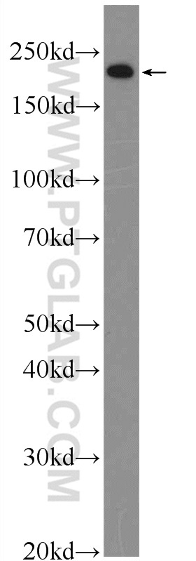
- Experimental details
- The MYH3 antibody from Proteintech is a rabbit polyclonal antibody to a peptide of human MYH3. This antibody recognizes human, mouse antigen. The MYH3 antibody has been validated for the following applications: ELISA, WB analysis.