Explore
 Validate
 Learn
Learn Western blot
 Immunocytochemistry
Antibody data
- Antibody Data
- Antigen structure
- References [1]
- Comments [0]
- Validations
- Immunocytochemistry [6]
- Immunohistochemistry [2]
- Flow cytometry [2]
- Other assay [3]
Submit
Validation data
Reference
Comment
Report error
- Product number
- MA5-32331 - Provider product page

- Provider
- Invitrogen Antibodies
- Product name
- Cyclin H Recombinant Rabbit Monoclonal Antibody (SN20-48)
- Antibody type
- Monoclonal
- Antigen
- Recombinant full-length protein
- Description
- Recombinant rabbit monoclonal antibodies are produced using in vitro expression systems. The expression systems are developed by cloning in the specific antibody DNA sequences from immunoreactive rabbits. Then, individual clones are screened to select the best candidates for production. The advantages of using recombinant rabbit monoclonal antibodies include: better specificity and sensitivity, lot-to-lot consistency, animal origin-free formulations, and broader immunoreactivity to diverse targets due to larger rabbit immune repertoire.
- Reactivity
- Human, Mouse
- Host
- Rabbit
- Isotype
- IgG
- Antibody clone number
- SN20-48
- Vial size
- 100 μL
- Concentration
- 1 mg/mL
- Storage
- Store at 4°C short term. For long term storage, store at -20°C, avoiding freeze/thaw cycles.
Submitted references Cyclin H predicts the poor prognosis and promotes the proliferation of ovarian cancer.
Peng C, Yang Y, Ji L, Yang P, Yang X, Zhang Y
Cancer cell international 2020;20:316
Cancer cell international 2020;20:316
No comments: Submit comment
Supportive validation
- Submitted by
- Invitrogen Antibodies (provider)
- Main image

- Experimental details
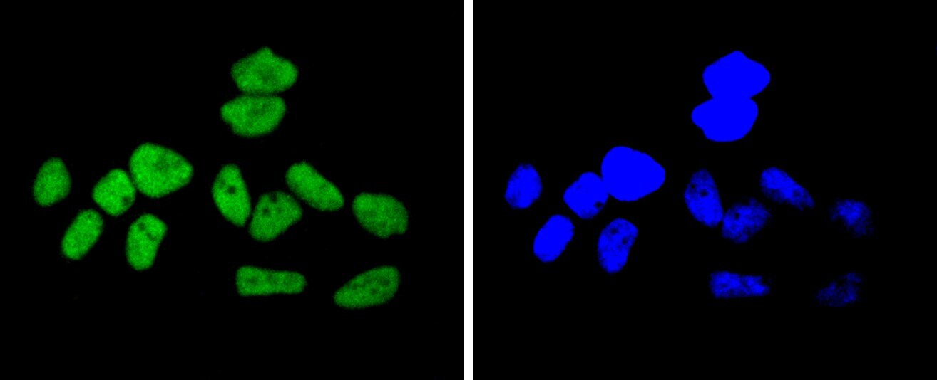
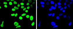
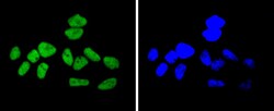
- Immunocytochemical analysis of Cyclin H in Hela cells using a Cyclin H Monoclonal antibody (Product # MA5-32331) as seen in green. The nuclear counter stain is DAPI (blue). Cells were fixed in paraformaldehyde, permeabilised with 0.25% Triton X100/PBS.
- Submitted by
- Invitrogen Antibodies (provider)
- Main image
- Experimental details
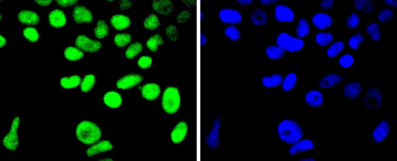
- Immunocytochemical analysis of Cyclin H in MCF-7 cells using a Cyclin H Monoclonal antibody (Product # MA5-32331) as seen in green. The nuclear counter stain is DAPI (blue). Cells were fixed in paraformaldehyde, permeabilised with 0.25% Triton X100/PBS.

- Submitted by
- Invitrogen Antibodies (provider)
- Main image

- Experimental details
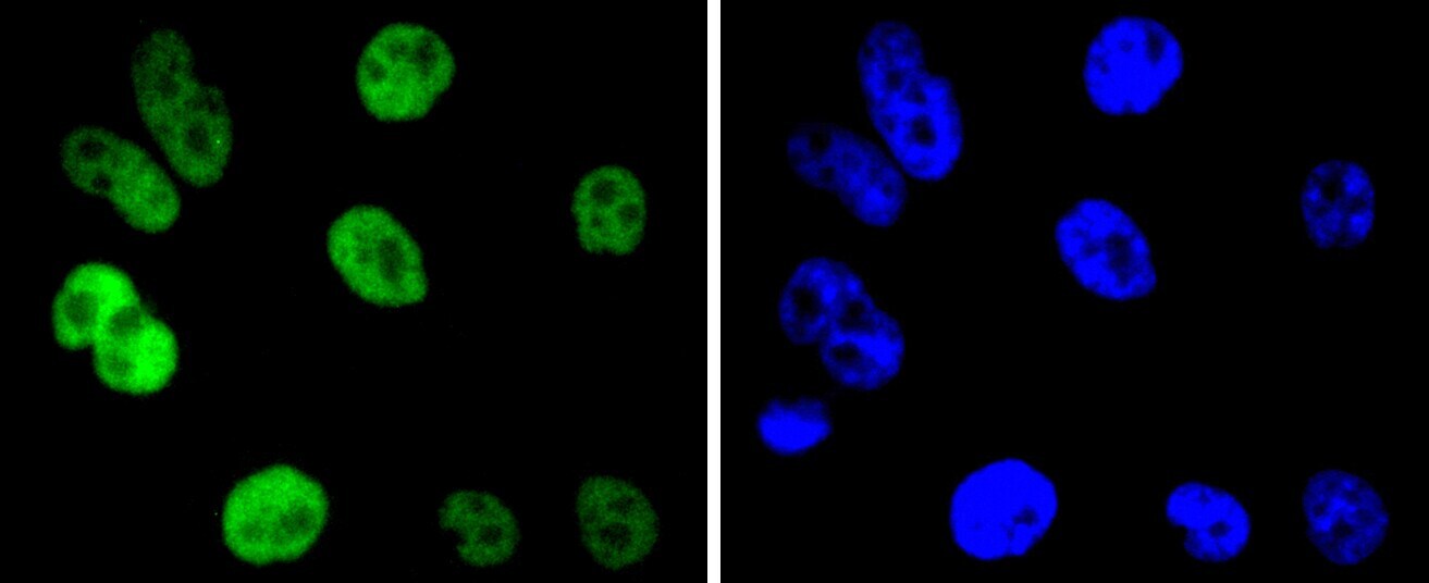
- Immunocytochemical analysis of Cyclin H in PC-3M cells using a Cyclin H Monoclonal antibody (Product # MA5-32331) as seen in green. The nuclear counter stain is DAPI (blue). Cells were fixed in paraformaldehyde, permeabilised with 0.25% Triton X100/PBS.

- Submitted by
- Invitrogen Antibodies (provider)
- Main image
- Experimental details
- Immunocytochemical analysis of Cyclin H in Hela cells using a Cyclin H Monoclonal antibody (Product # MA5-32331) as seen in green. The nuclear counter stain is DAPI (blue). Cells were fixed in paraformaldehyde, permeabilised with 0.25% Triton X100/PBS.

- Submitted by
- Invitrogen Antibodies (provider)
- Main image

- Experimental details
- Immunocytochemical analysis of Cyclin H in MCF-7 cells using a Cyclin H Monoclonal antibody (Product # MA5-32331) as seen in green. The nuclear counter stain is DAPI (blue). Cells were fixed in paraformaldehyde, permeabilised with 0.25% Triton X100/PBS.
- Submitted by
- Invitrogen Antibodies (provider)
- Main image
- Experimental details
- Immunocytochemical analysis of Cyclin H in PC-3M cells using a Cyclin H Monoclonal antibody (Product # MA5-32331) as seen in green. The nuclear counter stain is DAPI (blue). Cells were fixed in paraformaldehyde, permeabilised with 0.25% Triton X100/PBS.
Supportive validation
- Submitted by
- Invitrogen Antibodies (provider)
- Main image
- Experimental details
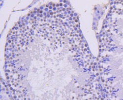
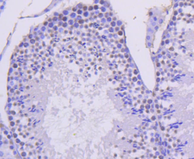
- Immunohistochemical analysis of Cyclin H of paraffin-embedded Mouse testis tissue using a Cyclin-H Monoclonal antibody (Product #MA5-32331). Counter stained with hematoxylin.
- Submitted by
- Invitrogen Antibodies (provider)
- Main image

- Experimental details
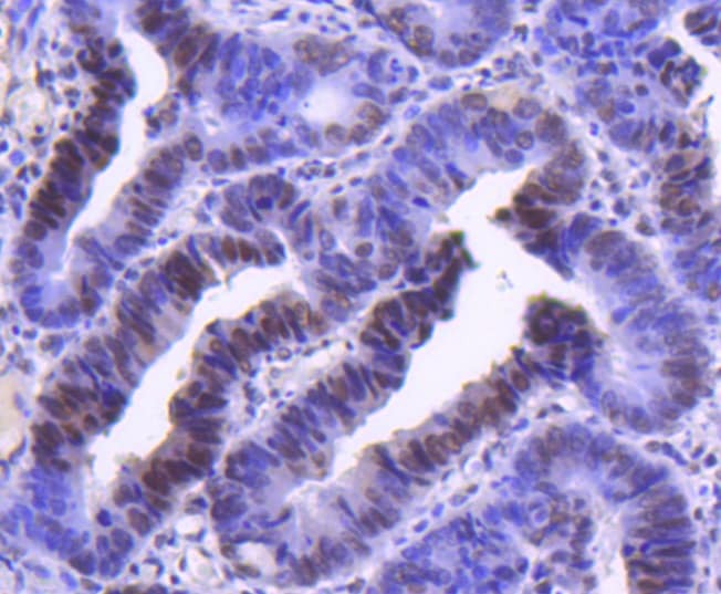
- Immunohistochemical analysis of Cyclin H of paraffin-embedded Human colon cancer tissue using a Cyclin-H Monoclonal antibody (Product #MA5-32331). Counter stained with hematoxylin.
Supportive validation
- Submitted by
- Invitrogen Antibodies (provider)
- Main image

- Experimental details
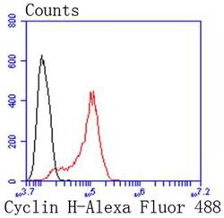
- Flow Cytometric analysis of Cyclin H in Hela cells using a Cyclin H Monoclonal Antibody (Product # MA5-32331) at a dilution of 1:50, as seen in red compared with an unlabelled control (cells without incubation with primary antibody; black). Alexa Fluor 488-conjugated goat anti rabbit IgG was used as the secondary antibody.

- Submitted by
- Invitrogen Antibodies (provider)
- Main image
- Experimental details
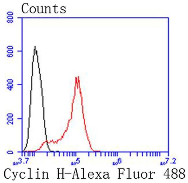
- Flow Cytometric analysis of Cyclin H in Hela cells using a Cyclin H Monoclonal Antibody (Product # MA5-32331) at a dilution of 1:50, as seen in red compared with an unlabelled control (cells without incubation with primary antibody; black). Alexa Fluor 488-conjugated goat anti rabbit IgG was used as the secondary antibody.
Supportive validation
- Submitted by
- Invitrogen Antibodies (provider)
- Main image
- Experimental details
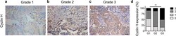
- Fig. 1 Immunohistochemistry staining of Cyclin H in ovarian cancer tissues. The expression of Cyclin H in ovarian cancer of grade 1 ( a ), grade 2 ( b ), and grade 3 ( c ) was measured by immunohistochemistry staining, and representative images are displayed. Scale bar, 20 um. d Quantification of Cyclin H expression in ovarian cancer (P < 0.001)

- Submitted by
- Invitrogen Antibodies (provider)
- Main image
- Experimental details
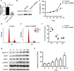
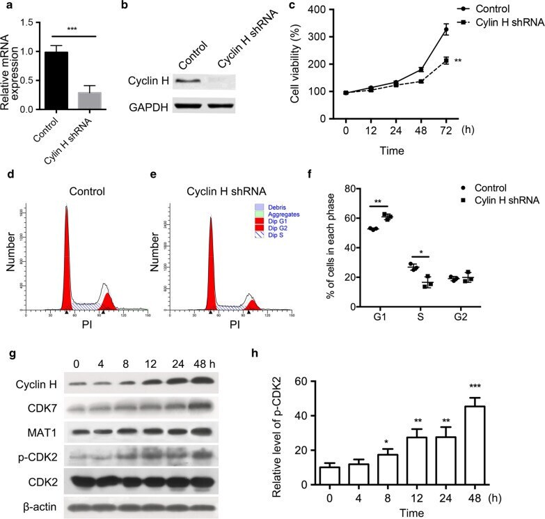
- Fig. 4 Cyclin H regulates the proliferation and cell cycle of ovarian cancer cells. a mRNA level of cyclin H in vector and cyclin H shRNA transfected ovarian cancer HO8910 cells. b The protein expression of cyclin H was detected by Western blot. c Proliferation difference between control and cyclin H silencing HO8910 cells. PI staining was performed to measure the percentage of cells in each cell cycle phase using Modfit software ( d and e ). f Quantification of the percentage cells in each cell cycle phase after transfection with cyclin H shRNA. g Expression of cyclin H, CDK7, MAT1, p-CDK2, and CDK2 in HO8910 cells after serum deprivation and refeeding. Serum-starved HO8910 cells were cultured in serum-containing medium for 4, 8, 12, 24, and 48 h, and cell lysates were analyzed by western blot. h Relative level of p-CDK2 was normalized to the total level of CDK2 at each time point. * P < 0.05, **P < 0.01, ***P < 0.001
- Submitted by
- Invitrogen Antibodies (provider)
- Main image

- Experimental details
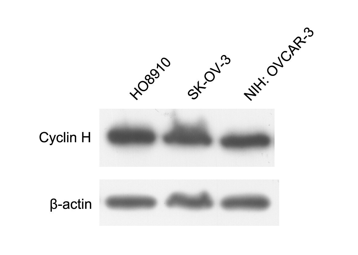
- Additional file 1: Figure S1. Expression of cyclin H in different ovarian cancer cell lines. The protein levels of cyclin H in HO8910, SK-OV-3, and NIH: OVCAR-3 cells were detected by western blot. Cyclin H was highly expressed in HO8910 cells.