Explore
 Validate
 Learn
LearnMA1-06503
antibody from Invitrogen Antibodies
Targeting: MUC1
ADMCKD, ADMCKD1, CD227, MCD, MCKD, MCKD1, PEM, PUM
 Western blot
 Immunohistochemistry
Antibody data
- Antibody Data
- Antigen structure
- References [13]
- Comments [0]
- Validations
- Western blot [1]
- Immunocytochemistry [1]
- Other assay [1]
Submit
Validation data
Reference
Comment
Report error
- Product number
- MA1-06503 - Provider product page

- Provider
- Invitrogen Antibodies
- Product name
- MUC1 Monoclonal Antibody (GP1.4)
- Antibody type
- Monoclonal
- Antigen
- Other
- Description
- MA1-06503 detects episialin in human samples. MA1-06503 has sucessfully been used in Western blotting, flow cytometry, immunocytochemistry, and immunohistochemistry. The MA1-06503 immunogen is a human milk fat globule. Store at 4ºC or in small aliquots at -20ºC if preferred. Avoid freeze-thaw cycles.
- Reactivity
- Human
- Host
- Mouse
- Isotype
- IgG
- Antibody clone number
- GP1.4
- Vial size
- 100 µg
- Concentration
- 1 mg/mL
- Storage
- Store at 4°C short term. For long term storage, store at -20°C, avoiding freeze/thaw cycles.
Submitted references Membrane-Tethered MUC1 Mucin Counter-Regulates the Phagocytic Activity of Macrophages.
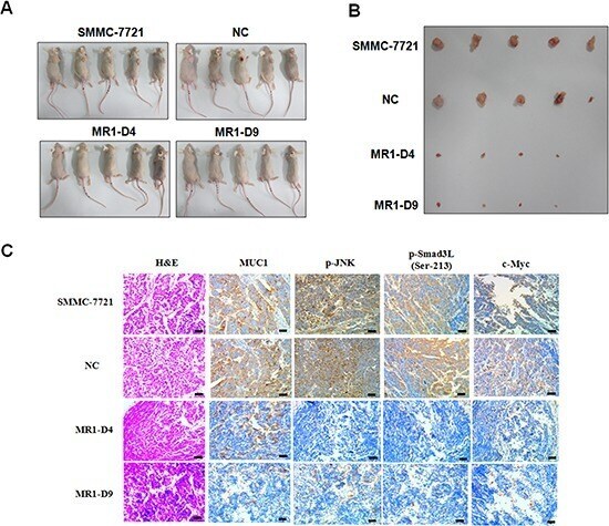
Mucin1 shifts Smad3 signaling from the tumor-suppressive pSmad3C/p21(WAF1) pathway to the oncogenic pSmad3L/c-Myc pathway by activating JNK in human hepatocellular carcinoma cells.
Expression of mesothelioma-related markers in meningiomas: an immunohistochemical study.
Multiple oncocytic cystadenoma with intraluminal crystalloids in parotid gland: case report.
Expression of human full-length MUC1 inhibits the proliferation and migration of a B16 mouse melanoma cell line.
Multinodular reticular schwannoma in the head and neck region: a potential diagnostic pitfall.
Anti-MUC1 antibody inhibits EGF receptor signaling in cancer cells.
Human embryonic stem cell-derived keratinocytes exhibit an epidermal transcription program and undergo epithelial morphogenesis in engineered tissue constructs.
Secondary involvement of the breast in T-cell non-Hodgkin lymphoma, an unusual example mimicking inflammatory breast carcinoma.
Myointimoma of the glans penis.
Pulmonary large cell carcinoma with rhabdoid phenotype.
Selective secretion and replenishment of discrete mucin glycoforms from intestinal goblet cells.
Selective secretion and replenishment of discrete mucin glycoforms from intestinal goblet cells.
Kato K, Uchino R, Lillehoj EP, Knox K, Lin Y, Kim KC
American journal of respiratory cell and molecular biology 2016 Apr;54(4):515-23
American journal of respiratory cell and molecular biology 2016 Apr;54(4):515-23
Mucin1 shifts Smad3 signaling from the tumor-suppressive pSmad3C/p21(WAF1) pathway to the oncogenic pSmad3L/c-Myc pathway by activating JNK in human hepatocellular carcinoma cells.
Li Q, Liu G, Yuan H, Wang J, Guo Y, Chen T, Zhai R, Shao D, Ni W, Tai G
Oncotarget 2015 Feb 28;6(6):4253-65
Oncotarget 2015 Feb 28;6(6):4253-65
Expression of mesothelioma-related markers in meningiomas: an immunohistochemical study.
Abdelzaher E, Abdallah DM
BioMed research international 2014;2014:968794
BioMed research international 2014;2014:968794
Multiple oncocytic cystadenoma with intraluminal crystalloids in parotid gland: case report.
Başak K, Kroğlu K
Medicine 2014 Dec;93(27):e246
Medicine 2014 Dec;93(27):e246
Expression of human full-length MUC1 inhibits the proliferation and migration of a B16 mouse melanoma cell line.
Wang F, Li Q, Ni W, Fang F, Sun X, Xie F, Wang J, Wang F, Gao S, Tai G
Oncology reports 2013 Jul;30(1):260-8
Oncology reports 2013 Jul;30(1):260-8
Multinodular reticular schwannoma in the head and neck region: a potential diagnostic pitfall.
Lau PP, Yau DT, Lau WH, Mak LS, Chan JK
International journal of surgical pathology 2013 Feb;21(1):54-8
International journal of surgical pathology 2013 Feb;21(1):54-8
Anti-MUC1 antibody inhibits EGF receptor signaling in cancer cells.
Hisatsune A, Nakayama H, Kawasaki M, Horie I, Miyata T, Isohama Y, Kim KC, Katsuki H
Biochemical and biophysical research communications 2011 Feb 18;405(3):377-81
Biochemical and biophysical research communications 2011 Feb 18;405(3):377-81
Human embryonic stem cell-derived keratinocytes exhibit an epidermal transcription program and undergo epithelial morphogenesis in engineered tissue constructs.
Metallo CM, Azarin SM, Moses LE, Ji L, de Pablo JJ, Palecek SP
Tissue engineering. Part A 2010 Jan;16(1):213-23
Tissue engineering. Part A 2010 Jan;16(1):213-23
Secondary involvement of the breast in T-cell non-Hodgkin lymphoma, an unusual example mimicking inflammatory breast carcinoma.
Kelten C, Kabukcu S, Sen N, Teke Z, Yaren A, Erdem E, Duzcan E
Archives of gynecology and obstetrics 2009 Jul;280(1):149-52
Archives of gynecology and obstetrics 2009 Jul;280(1):149-52
Myointimoma of the glans penis.
Vardar E, Gunlusoy B, Arslan M, Kececi S
Pathology international 2007 Mar;57(3):158-61
Pathology international 2007 Mar;57(3):158-61
Pulmonary large cell carcinoma with rhabdoid phenotype.
Yilmazbayhan D, Ates LE, Dilege S, Gulluoglu M, Tanju S, Kalayci G
Annals of diagnostic pathology 2005 Aug;9(4):223-6
Annals of diagnostic pathology 2005 Aug;9(4):223-6
Selective secretion and replenishment of discrete mucin glycoforms from intestinal goblet cells.
Stanley CM, Phillips TE
The American journal of physiology 1999 Jul;277(1):G191-200
The American journal of physiology 1999 Jul;277(1):G191-200
Selective secretion and replenishment of discrete mucin glycoforms from intestinal goblet cells.
Stanley CM, Phillips TE
The American journal of physiology 1999 Jul;277(1 Pt 1):G191-200
The American journal of physiology 1999 Jul;277(1 Pt 1):G191-200
No comments: Submit comment
Supportive validation
- Submitted by
- Invitrogen Antibodies (provider)
- Main image

- Experimental details
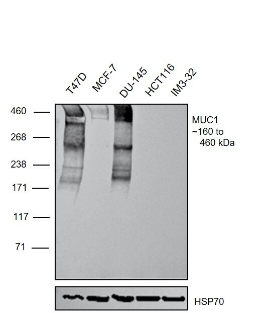
- Western blot was performed using Anti-MUC1 Monoclonal Antibody (GP1.4)(Product # MA1-06503) and a ~180, 260 and 460kDa band corresponding to MUC1 was observed across T47D, MCF-7 and DU-145 compared to HCT-116 and IMR-32. Membrane enriched extracts (50 µg lysate) of T-47D (Lane 1), MCF7 (Lane 2), DU 145 (Lane 3), HCT 116 (Lane 4), IMR-32 (Lane 5) were electrophoresed using NuPAGE™ 3-8% Tris-Acetate Protein Gel (Product # EA0378BOX). Resolved proteins were then transferred onto a Nitrocellulose membrane (Product # LC2002) by iBlot® 2 Dry Blotting System (Product # IB21001). The blot was probed with the primary antibody (1:1000) and detected by chemiluminescence with Goat anti-Mouse IgG (H+L) Superclonal™ Recombinant Secondary Antibody, HRP (Product # A28177,1:4000) using the iBright FL 1000 (Product # A32752). Chemiluminescent detection was performed using SuperSignal™ West Atto Ultimate Sensitivity Substrate (Product # A38556). A streak like pattern was observed in the positive cell lines
Supportive validation
- Submitted by
- Invitrogen Antibodies (provider)
- Main image
- Experimental details
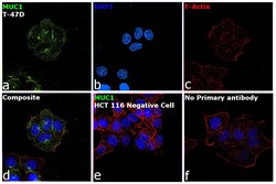
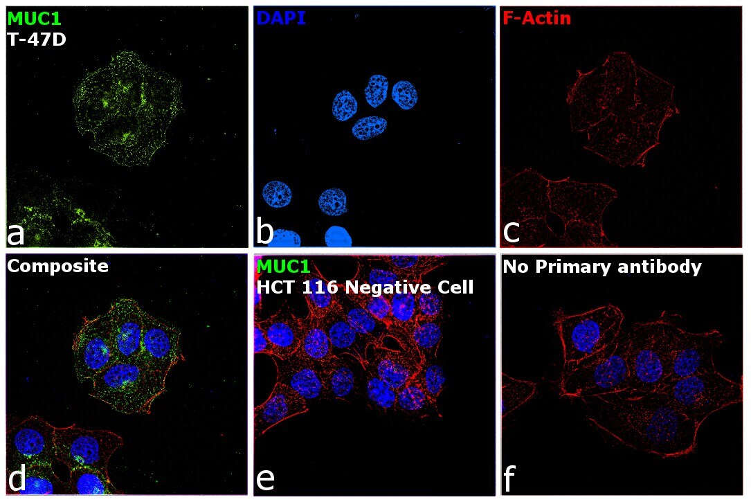
- Immunofluorescence analysis of MUC1 was performed using 70% confluent log phase T-47D cells. The cells were fixed with 4% paraformaldehyde for 10 minutes, permeabilized with 0.1% Triton™ X-100 for 15 minutes, and blocked with 2% BSA for 45 minutes at room temperature. The cells were labeled with MUC1 Monoclonal Antibody (GP1.4) (Product # MA1-06503) at 1:100 dilution in 0.1% BSA, incubated at 4 degree celsius overnight and then labeled with Donkey anti-Mouse IgG (H+L) Highly Cross-Adsorbed Secondary Antibody, Alexa Fluor Plus 488 (Product # A32766), at 1:2000 dilution, for 45 minutes at room temperature (Panel a: Green). Nuclei (Panel b: Blue) were stained with ProLong™ Diamond Antifade Mountant with DAPI (Product # P36962). F-actin (Panel c: Red) was stained with Rhodamine Phalloidin (Product # R415, 1:300). Panel d represents the merged image showing Plasma membrane and cytoplasm localization. Panel e represents HCT116. Panel f represents control cells with no primary antibody to assess background. The images were captured at 60X magnification.
Supportive validation
- Submitted by
- Invitrogen Antibodies (provider)
- Main image
- Experimental details
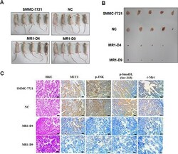
- Figure 5 Knockdown of MUC1 suppresses tumor growth and JNK/pSmad3L/c-Myc pathway in mice (A) BALB/c nude mice subcutaneous transplant tumor model was established using SMMC-7721, NC, MR1-D4 and MR1-D9 cells. (B) Tumors in mice were dissected and photographed on day 21 post-injection. The images were captured showing tumor sizes for each group. (C) Tumors from mice were detected for the expression of MUC1, p-JNK, p-Smad3L (Ser-213) and c-Myc by immunohistochemical staining. Sections were examined on an inverted fluorescence microscope (IX71; Olympus) at 100 x magnification. The scale bar indicates 50 mum.