 Explore
 Validate
 Learn
LearnMAB6298
antibody from Novus Biologicals
Targeting: MUC1
ADMCKD, ADMCKD1, CD227, MCD, MCKD, MCKD1, PEM, PUM
 Western blot
 Immunocytochemistry
Antibody data
- Antibody Data
- Antigen structure
- References [1]
- Comments [0]
- Validations
- Western blot [2]
- Immunohistochemistry [1]
- Flow cytometry [1]
Submit
Validation data
Reference
Comment
Report error
- Product number
- MAB6298 - Provider product page

- Provider
- Novus Biologicals
- Product name
- Mouse Monoclonal MUC1 Antibody
- Antibody type
- Monoclonal
- Description
- Protein A or G purified from hybridoma culture supernatant. Detects human MUC-1 in direct ELISAs and Western blots. In direct ELISAs, no cross-reactivity with recombinant human (rh) MUC-20, rhMUC-20S, or rhCA125/MUC-16 was observed.
- Reactivity
- Human
- Host
- Mouse
- Conjugate
- Unconjugated
- Isotype
- IgG
- Vial size
- 100 ug
- Concentration
- LYOPH
- Storage
- Use a manual defrost freezer and avoid repeated freeze-thaw cycles. 12 months from date of receipt, -20 to -70 degreesC as supplied. 1 month, 2 to 8 degreesC under sterile conditions after reconstitution. 6 months, -20 to -70 degreesC under sterile conditions after reconstitution.
Submitted references IL4 Primes the Dynamics of Breast Cancer Progression via DUSP4 Inhibition.
Gaggianesi M, Turdo A, Chinnici A, Lipari E, Apuzzo T, Benfante A, Sperduti I, Di Franco S, Meraviglia S, Lo Presti E, Dieli F, Caputo V, Militello G, Vieni S, Stassi G, Todaro M
Cancer research 2017 Jun 15;77(12):3268-3279
Cancer research 2017 Jun 15;77(12):3268-3279
No comments: Submit comment
Supportive validation
- Submitted by
- Novus Biologicals (provider)
- Main image

- Experimental details
- Detection of Human MUC-1 by Western Blot. Western blot shows lysates of human lung tissue. PVDF Membrane was probed with 2 µg/mL of Mouse Anti-Human MUC-1 Monoclonal Antibody (Catalog # MAB6298) followed by HRP-conjugated Anti-Mouse IgG Secondary Antibody (Catalog # HAF007). A specific band was detected for MUC-1 at approximately 300 kDa (as indicated). This experiment was conducted under non-reducing conditions and using Immunoblot Buffer Group 1.

- Submitted by
- Novus Biologicals (provider)
- Main image

- Experimental details
- Detection of Human MUC-1 by Simple WesternTM. Simple Western lane view shows lysates of human lung tissue, loaded at 0.2 mg/mL. Specific bands were detected for MUC-1 at approximately 300-500 kDa (as indicated) using 40 µg/mL of Mouse Anti-Human MUC-1 Monoclonal Antibody (Catalog # MAB6298). This experiment was conducted under reducing conditions and using the 66-440 kDa separation system.

Supportive validation
- Submitted by
- Novus Biologicals (provider)
- Main image

- Experimental details
- MUC-1 in Human Breast. MUC-1 was detected in immersion fixed paraffin-embedded sections of human breast using Mouse Anti-Human MUC-1 Monoclonal Antibody (Catalog # MAB6298) at 15 µg/mL overnight at 4 °C. Before incubation with the primary antibody, tissue was subjected to heat-induced epitope retrieval using Antigen Retrieval Reagent-Basic (Catalog # CTS013). Tissue was stained using the Anti-Mouse HRP-DAB Cell & Tissue Staining Kit (brown; Catalog # CTS002) and counterstained with hematoxylin (blue). Specific staining was localized to cytoplasm of epithelial cells. View our protocol for Chromogenic IHC Staining of Paraffin-embedded Tissue Sections.

Supportive validation
- Submitted by
- Novus Biologicals (provider)
- Main image
- Experimental details
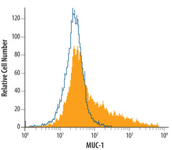
- Detection of MUC-1 in MCF-7 Human Cell Line by Flow Cytometry. MCF-7 human breast cancer cell line was stained with Mouse Anti-Human MUC-1 Monoclonal Antibody (Catalog # MAB6298, filled histogram) or isotype control antibody (Catalog # MAB0041, open histogram), followed by Allophycocyanin-conjugated Anti-Mouse IgG F(ab')2 Secondary Antibody (Catalog # F0101B).
