 Explore
 Validate
 Learn
LearnNBP2-62565
antibody from Novus Biologicals
Targeting: MUC1
ADMCKD, ADMCKD1, CD227, MCD, MCKD, MCKD1, PEM, PUM
 Western blot
 ELISA
ELISAAntibody data
- Antibody Data
- Antigen structure
- References [0]
- Comments [0]
- Validations
- Western blot [1]
- Immunohistochemistry [1]
- Flow cytometry [1]
Submit
Validation data
Reference
Comment
Report error
- Product number
- NBP2-62565 - Provider product page

- Provider
- Novus Biologicals
- Product name
- Rabbit Monoclonal MUC1 Antibody
- Antibody type
- Monoclonal
- Description
- Protein A purified. This antibody recognises MUC1, a tumor associated antigen which is expressed in >90% ovarian carcinomas. This antigen is a high molecular weight (M, 80,000-200,000) glycoprotein.
- Reactivity
- Human
- Host
- Rabbit
- Isotype
- IgG
- Vial size
- 0.2 mg
- Storage
- Store at 4C for up to 3 months. For longer storage, aliquot and store at -20C.
No comments: Submit comment
Supportive validation
- Submitted by
- Novus Biologicals (provider)
- Main image
- Experimental details
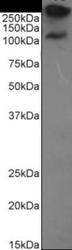
- Western Blot: MUC-1 Antibody (HMFG2) [NBP2-62565] - Western Blot using anti-MUC1 antibody HMFG2. MCF-7 cell lysate (35ug protein in RIPA buffer) were resolved on a 10% SDS PAGE gel and blots probed with the chimeric rabbit version of HMFG2 (NBP2-62565) at 0.1 ug/ml before detection using an anti-rabbit secondary antibody. A primary incubation of 1h was used and protein was detected by chemiluminescence. The predicted band size for unmodified MUC1 is 122.1kDa, though in breast cancer cell lines like MCF-7 MUC1 can be up to 90% glycosylated (c.f. Mueller et al. PMID: 10373415; T47D cells) and expected band sizes are ~250-300kDa. Thus the two bands likely represent processed (>250kDa) and unprocessed (~121kDa) populations of the protein. NBP2-62565 successfully detected human MUC1 in MCF-7 breast cancer cells.

Supportive validation
- Submitted by
- Novus Biologicals (provider)
- Main image
- Experimental details
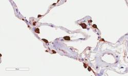
- Immunohistochemistry-Paraffin: MUC-1 Antibody (HMFG2) [NBP2-62565] - Immunohistochemical staining of human lung tissue using anti-MUC1 antibody. HMFG2 Anti-MUC1 (Mucin-1) staining of paraffin embedded human lung tissue using the rabbit-chimeric version of HMFG2 (NBP2-62565). Antigen retreival was acheived by microwaving in citrate buffer (pH6), followed by blocking with protein block serum-free buffer. Primary antibody incubation with NBP2-62565 was carried out at 4 ug/ml for 30 minutes. Samples were then incubated with an anti-rabbit IgG HRP secondary antibody for 20 mins followed by DAB (3,3'-diaminobenzidine), and counter-staining with haemotoxylin. Strong staining of type II pneumocytes may be observed. Recommended concentration, 1-2 ug/ml.

Supportive validation
- Submitted by
- Novus Biologicals (provider)
- Main image

- Experimental details
- Flow Cytometry: MUC-1 Antibody (HMFG2) [NBP2-62565] - Flow-cytometry using the anti-MUC1 antibody HMFG2. MCF-7 cells were stained with unimmunized rabbit IgG antibody (black line) or the rabbit-chimeric version of HMFG2 (NBP2-62565, blue line) at a concentration of 10 ug/ml for 30 mins at RT. After washing, bound antibody was detected using an anti-rabbit IgG JK (FITC-conjugate) antibody at 2 ug/ml and cells analyzed on a FACSCanto flow-cytometer.
