Explore
 Validate
 Learn
LearnMA5-32265
antibody from Invitrogen Antibodies
Targeting: MUC1
ADMCKD, ADMCKD1, CD227, MCD, MCKD, MCKD1, PEM, PUM
 Western blot
Antibody data
- Antibody Data
- Antigen structure
- References [1]
- Comments [0]
- Validations
- Western blot [3]
- Immunocytochemistry [4]
- Immunohistochemistry [5]
- Flow cytometry [1]
- Other assay [1]
Submit
Validation data
Reference
Comment
Report error
- Product number
- MA5-32265 - Provider product page

- Provider
- Invitrogen Antibodies
- Product name
- MUC1 Recombinant Rabbit Monoclonal Antibody (SN06-80)
- Antibody type
- Monoclonal
- Antigen
- Synthetic peptide
- Description
- Recombinant rabbit monoclonal antibodies are produced using in vitro expression systems. The expression systems are developed by cloning in the specific antibody DNA sequences from immunoreactive rabbits. Then, individual clones are screened to select the best candidates for production. The advantages of using recombinant rabbit monoclonal antibodies include: better specificity and sensitivity, lot-to-lot consistency, animal origin-free formulations, and broader immunoreactivity to diverse targets due to larger rabbit immune repertoire.
- Reactivity
- Human
- Host
- Rabbit
- Isotype
- IgG
- Antibody clone number
- SN06-80
- Vial size
- 100 μL
- Concentration
- 1 mg/mL
- Storage
- Store at 4°C short term. For long term storage, store at -20°C, avoiding freeze/thaw cycles.
Submitted references (89)Zr-Labeled AR20.5: A MUC1-Targeting ImmunoPET Probe.
Fung K, Vivier D, Keinänen O, Sarbisheh EK, Price EW, Zeglis BM
Molecules (Basel, Switzerland) 2020 May 15;25(10)
Molecules (Basel, Switzerland) 2020 May 15;25(10)
No comments: Submit comment
Supportive validation
- Submitted by
- Invitrogen Antibodies (provider)
- Main image
- Experimental details
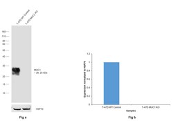
- Knockout of MUC1 was achieved by CRISPR-Cas9 genome editing using LentiArray™ Lentiviral sgRNA (Product # A32042, Assay ID CRISPR654939_LV) and LentiArray Cas9 Lentivirus (Product # A32064). Western blot analysis of MUC1 was performed by loading 30 µg of T-47D Wild Type (Lane 1) and T-47D MUC1 KO (Lane 2) whole cell extracts. The samples were electrophoresed using NuPAGE™ Novex™ 4-12% Bis-Tris Protein Gel (Product # NP0321BOX). Resolved proteins were then transferred onto a nitrocellulose membrane (Product # IB23001) by iBlot® 2 Dry Blotting System (Product # IB21001). The blot was probed with MUC1 Recombinant Rabbit Monoclonal Antibody (SN06-80) (Product # MA5-32265, 1:1,000) and detected by Goat anti-Rabbit IgG (H+L) Superclonal™ Recombinant Secondary Antibody, HRP (Product # A27036, 1:10,000) using the iBright™ FL1500 (Product # A44115). Chemiluminescent detection was performed using SuperSignal™ West Dura Extended Duration Substrate (Product # 34076). Loss of signal upon CRISPR mediated knockout (KO) using the LentiArray™ CRISPR product line confirms that antibody is specific to MUC1.
- Submitted by
- Invitrogen Antibodies (provider)
- Main image
- Experimental details
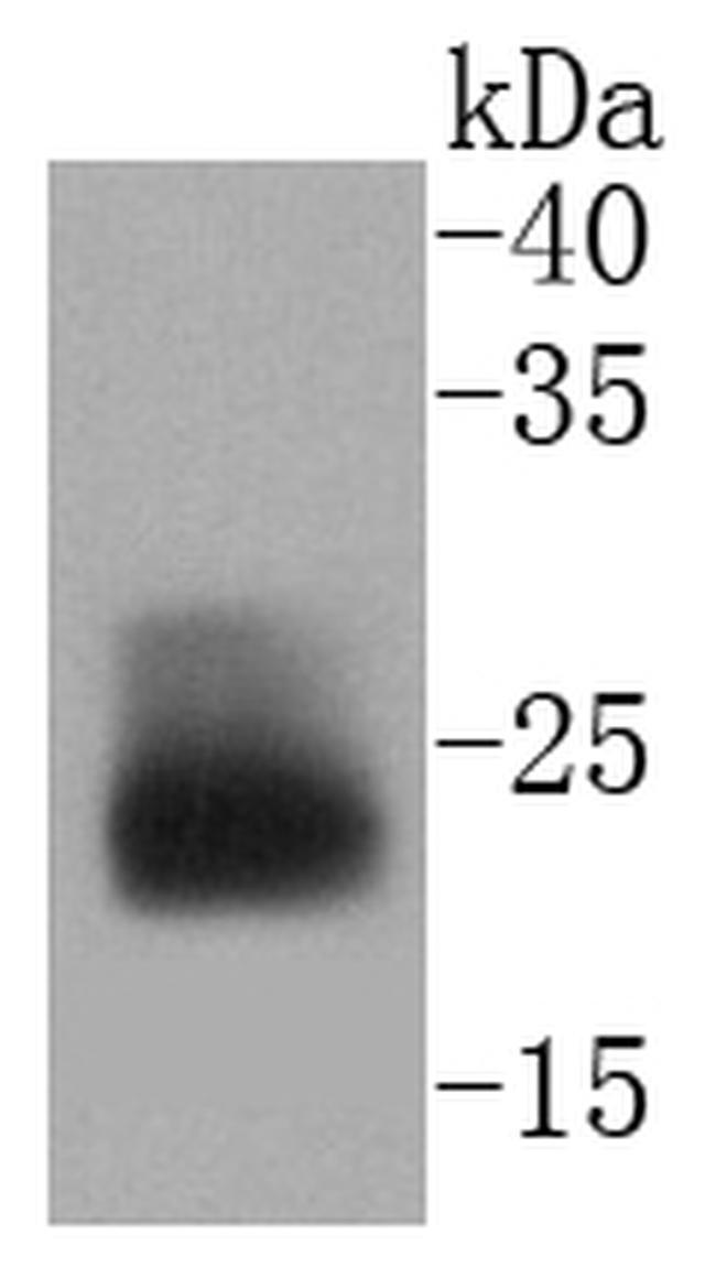
- Western blot analysis of MUC1 in Hela cell lysate using a MUC1 Monoclonal antibody (Product # MA5-32265) at a dilution of 1:1,000.
- Submitted by
- Invitrogen Antibodies (provider)
- Main image
- Experimental details
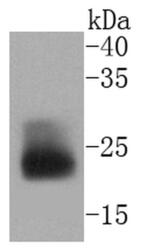
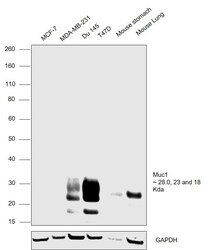
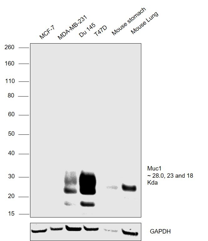
- Western blot analysis was performed on whole cell extracts (30 µg lysate) of MCF-7 (Lane 1), MDA-MB-231 (Lane 2), DU 145 (Lane 3), T47D (Lane 4), Mouse Stomach (Lane 5) and Mouse Lung (Lane 6). Bands at ~28, ~23 and ~18 kDa were observed corresponding to MUC1 across all the cell lines tested except MCF-7 and MDA-MB-231 which are reported to be negative. The blot was probed with Anti-MUC1 Recombinant Rabbit Monoclonal Antibody (Product # MA5-32265, 1:1,000 dilution). Resolved proteins were then transferred onto a nitrocellulose membrane (Product # IB23001) by iBlot® 2 Dry Blotting System (Product # IB21001) and detected by chemiluminescence using Goat anti-Rabbit IgG (Heavy Chain) Superclonal™ Recombinant Secondary Antibody, HRP conjugate (Product # A27036, 0.25 µg/mL, 1:4,000 dilution) using the iBright FL 1000 (Product # A32752). Chemiluminescent detection was performed using Novex® ECL Chemiluminescent Substrate Reagent Kit (Product # WP20005).
Supportive validation
- Submitted by
- Invitrogen Antibodies (provider)
- Main image

- Experimental details
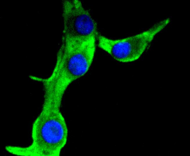
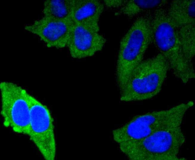
- Immunocytochemical analysis of MUC1 in Hela cells using a MUC1 Monoclonal antibody (Product # MA5-32265) as seen in green. The nuclear counter stain is DAPI (blue). Cells were fixed in paraformaldehyde, permeabilised with 0.25% Triton X100/PBS.

- Submitted by
- Invitrogen Antibodies (provider)
- Main image

- Experimental details
- Immunocytochemical analysis of MUC1 in B16-F1 cells using a MUC1 Monoclonal antibody (Product # MA5-32265) as seen in green. The nuclear counter stain is DAPI (blue). Cells were fixed in paraformaldehyde, permeabilised with 0.25% Triton X100/PBS.
- Submitted by
- Invitrogen Antibodies (provider)
- Main image

- Experimental details
- Immunocytochemical analysis of MUC1 in Hela cells using a MUC1 Monoclonal antibody (Product # MA5-32265) as seen in green. The nuclear counter stain is DAPI (blue). Cells were fixed in paraformaldehyde, permeabilised with 0.25% Triton X100/PBS.
- Submitted by
- Invitrogen Antibodies (provider)
- Main image

- Experimental details
- Immunocytochemical analysis of MUC1 in B16-F1 cells using a MUC1 Monoclonal antibody (Product # MA5-32265) as seen in green. The nuclear counter stain is DAPI (blue). Cells were fixed in paraformaldehyde, permeabilised with 0.25% Triton X100/PBS.

Supportive validation
- Submitted by
- Invitrogen Antibodies (provider)
- Main image

- Experimental details
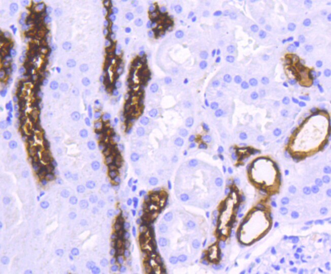
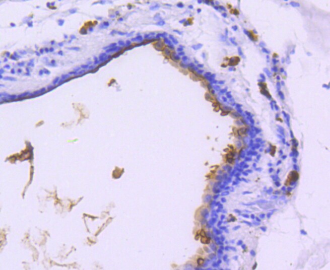
- Immunohistochemical analysis of MUC1 of paraffin-embedded Human kidney tissue using a MUC1 Monoclonal antibody (Product #MA5-32265). Counter stained with hematoxylin.
- Submitted by
- Invitrogen Antibodies (provider)
- Main image

- Experimental details
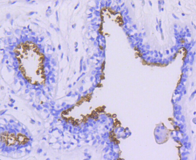
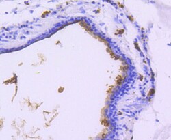
- Immunohistochemical analysis of MUC1 of paraffin-embedded Human breast tissue using a MUC1 Monoclonal antibody (Product #MA5-32265). Counter stained with hematoxylin.
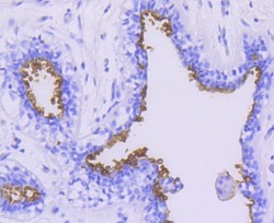
- Submitted by
- Invitrogen Antibodies (provider)
- Main image
- Experimental details
- Immunohistochemical analysis of MUC1 of paraffin-embedded Human breast carcinoma tissue using a MUC1 Monoclonal antibody (Product #MA5-32265). Counter stained with hematoxylin.
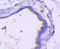
- Submitted by
- Invitrogen Antibodies (provider)
- Main image
- Experimental details
- Immunohistochemical analysis of MUC1 of paraffin-embedded Human breast tissue using a MUC1 Monoclonal antibody (Product #MA5-32265). Counter stained with hematoxylin.

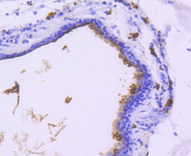
- Submitted by
- Invitrogen Antibodies (provider)
- Main image
- Experimental details
- Immunohistochemical analysis of MUC1 of paraffin-embedded Human breast carcinoma tissue using a MUC1 Monoclonal antibody (Product #MA5-32265). Counter stained with hematoxylin.
Supportive validation
- Submitted by
- Invitrogen Antibodies (provider)
- Main image
- Experimental details
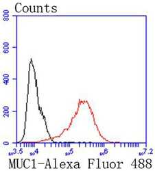
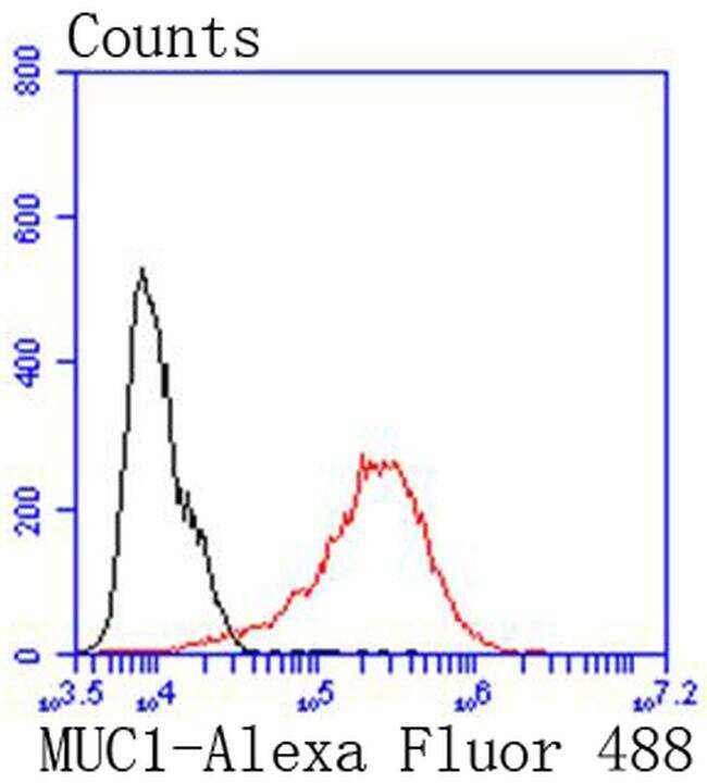
- Flow Cytometric analysis of MUC1 in MCF-7 cells using a MUC1 Monoclonal Antibody (Product # MA5-32265) at a dilution of 1:50, as seen in red compared with an unlabelled control (cells without incubation with primary antibody; black). Alexa Fluor 488-conjugated goat anti rabbit IgG was used as the secondary antibody.
Supportive validation
- Submitted by
- Invitrogen Antibodies (provider)
- Main image
- Experimental details
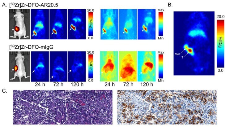
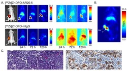
- Figure 4 ( A ) Bioluminescence images (left) as well as planar (center) and maximum intensity projection (right; scaled to a minimum of 0% and maximum of 100%) PET images of representative athymic nude mice bearing orthotopic SKOV3-Red-FLuc xenografts obtained 24, 72, and 120 h following the intravenous tail vein injection of [ 89 Zr]Zr-DFO-AR20.5 or [ 89 Zr]Zr-DFO-mIgG. The white arrows mark the tumors; ( B ) Planar PET image of a representative athymic nude mouse bearing an orthotopic SKOV3-Red-FLuc xenograft collected at 120 h post-injection of [ 89 Zr]Zr-DFO-AR20.5. The white arrows mark the tumor (T) and a peritoneal metastatic lesion (Met); ( C ) Hematoxylin and eosin staining (10x magnified; left) and immunohistochemical staining (10x magnified; right) of the peritoneal metastatic lesion from the representative mouse, with brown staining indicating the expression of MUC1.