Explore
 Validate
 Learn
Learn Western blot
 Immunocytochemistry
Antibody data
- Antibody Data
- Antigen structure
- References [0]
- Comments [0]
- Validations
- Immunocytochemistry [2]
- Flow cytometry [2]
Submit
Validation data
Reference
Comment
Report error
- Product number
- MA3-069 - Provider product page

- Provider
- Invitrogen Antibodies
- Product name
- TAF10 Monoclonal Antibody (23TA-1H8)
- Antibody type
- Monoclonal
- Antigen
- Synthetic peptide
- Description
- MA3-069 detects TAF10 from human, rat, and non-human primate samples. MA3-069 does not detect the TAF10 in mouse samples MA3-069 has been successfully used in Western blot, immunofluorescence and flow cytometry applications. The MA3-069 immunogen is the synthetic peptide coupled to ovalalbumin aa 1 to aa 20.
- Reactivity
- Human, Rat
- Host
- Mouse
- Isotype
- IgG
- Antibody clone number
- 23TA-1H8
- Vial size
- 50 μL
- Concentration
- Conc. Not Determined
- Storage
- -20°C, Avoid Freeze/Thaw Cycles
No comments: Submit comment
Supportive validation
- Submitted by
- Invitrogen Antibodies (provider)
- Main image
- Experimental details
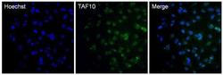
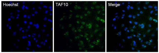
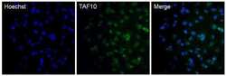
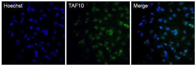
- IImmunofluorescent analysis of TAF10 in HepG2 cells. The cells were fixed with 4% paraformaldehyde in PBS for 15 minutes, permeabilized with 0.1% Triton X-100 for 15 minutes, and blocked with 3% BSA in PBS for 30 minutes at room temperature. Cells were stained with a TAF10 mouse monoclonal antibody (Product # MA3-069) at a dilution of 1:2000 in blocking buffer for 1 hour at room temperature, and then incubated with Goat anti-Mouse IgG (H+L) Superclonal Secondary Antibody, Alexa Fluor® 488 conjugate (Product # A28175) at a dilution of 1:1000 for at least 30 minutes at room temperature in the dark (green). Nuclei (blue) were stained with Hoechst 33342 (Product # 62249). Images were taken on a Thermo Scientific ToxInsight Instrument at 20X magnification.
- Submitted by
- Invitrogen Antibodies (provider)
- Main image
- Experimental details
- IImmunofluorescent analysis of TAF10 in HepG2 cells. The cells were fixed with 4% paraformaldehyde in PBS for 15 minutes, permeabilized with 0.1% Triton X-100 for 15 minutes, and blocked with 3% BSA in PBS for 30 minutes at room temperature. Cells were stained with a TAF10 mouse monoclonal antibody (Product # MA3-069) at a dilution of 1:2000 in blocking buffer for 1 hour at room temperature, and then incubated with Goat anti-Mouse IgG (H+L) Superclonal Secondary Antibody, Alexa Fluor® 488 conjugate (Product # A28175) at a dilution of 1:1000 for at least 30 minutes at room temperature in the dark (green). Nuclei (blue) were stained with Hoechst 33342 (Product # 62249). Images were taken on a Thermo Scientific ToxInsight Instrument at 20X magnification.
Supportive validation
- Submitted by
- Invitrogen Antibodies (provider)
- Main image
- Experimental details
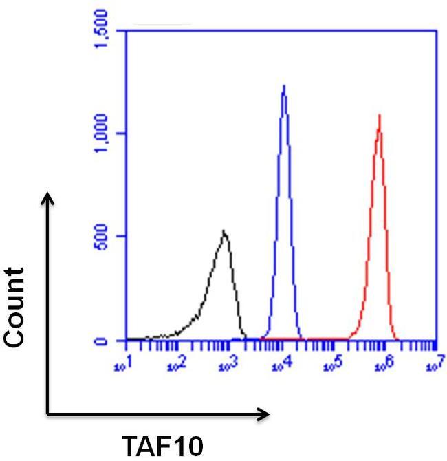
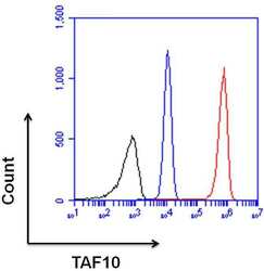
- Flow cytometry analysis of TAF10 was done on HeLa cells. Cells were fixed, permeabilized and stained with a TAF10 mouse monoclonal antibody (Product # MA3-069, red histogram) at a dilution of 1:100. After incubation of the primary antibody on ice for 1 hour, the cells were stained with a Goat anti-Mouse IgG (H+L) Secondary Antibody, DyLight 650 conjugate (Product # 84545) at a dilution of 1:50 for at least 30 minutes on ice. A representative 10,000 cells were acquired for each sample. The black histogram represents unstained control cells and the blue histogram represents no-primary-antibody control.
- Submitted by
- Invitrogen Antibodies (provider)
- Main image

- Experimental details
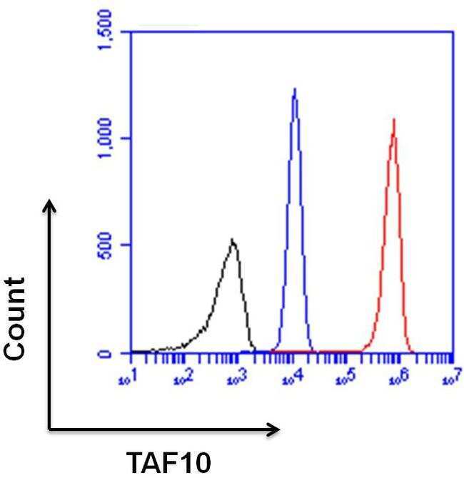
- Flow cytometry analysis of TAF10 was done on HeLa cells. Cells were fixed, permeabilized and stained with a TAF10 mouse monoclonal antibody (Product # MA3-069, red histogram) at a dilution of 1:100. After incubation of the primary antibody on ice for 1 hour, the cells were stained with a Goat anti-Mouse IgG (H+L) Secondary Antibody, DyLight 650 conjugate (Product # 84545) at a dilution of 1:50 for at least 30 minutes on ice. A representative 10,000 cells were acquired for each sample. The black histogram represents unstained control cells and the blue histogram represents no-primary-antibody control.