Antibody data
- Antibody Data
- Antigen structure
- References [1]
- Comments [0]
- Validations
- Western blot [1]
- Immunohistochemistry [11]
Submit
Validation data
Reference
Comment
Report error
- Product number
- HPA022856 - Provider product page
- Provider
- Atlas Antibodies
- Proper citation
- Atlas Antibodies Cat#HPA022856, RRID:AB_1858253
- Product name
- Anti-RIDA
- Antibody type
- Polyclonal
- Reactivity
- Human, Rat
- Host
- Rabbit
- Conjugate
- Unconjugated
- Antigen sequence
MSSLIRRVISTAKAPGAIGPYSQAVLVDRTIYISG
QIGMDPSSGQLVSGGVAEEAKQALKNMGEILKAA- Isotype
- IgG
- Vial size
- 100 µl
- Storage
- Store at +4°C for short term storage. Long time storage is recommended at -20°C.
Submitted references Y-box binding protein 1 and RNase UK114 mediate monocyte chemoattractant protein 1 mRNA stability in vascular smooth muscle cells.
Dhawan L, Liu B, Pytlak A, Kulshrestha S, Blaxall BC, Taubman MB
Molecular and cellular biology 2012 Sep;32(18):3768-75
No comments: Submit comment
Enhanced validation
- Submitted by
- Atlas Antibodies (provider)
- Enhanced method
- Independent antibody validation
- Main image
- Experimental details
- Western blot analysis using Anti-RIDA antibody HPA022856 (A) shows similar pattern to independent antibody HPA023489 (B).
Enhanced validation
Enhanced validation
Supportive validation
- Submitted by
- Atlas Antibodies (provider)
- Enhanced method
- Orthogonal validation
- Main image
- Experimental details
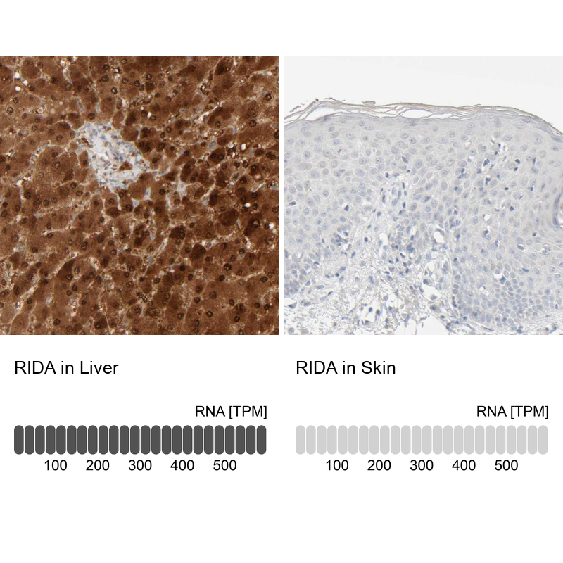
- Immunohistochemistry analysis in human liver and skin tissues using HPA022856 antibody. Corresponding RIDA RNA-seq data are presented for the same tissues.
- Sample type
- HUMAN
Enhanced validation
- Submitted by
- Atlas Antibodies (provider)
- Enhanced method
- Independent antibody validation
- Main image
- Experimental details
- Immunohistochemical staining of human fallopian tube, kidney, liver and skin using Anti-RIDA antibody HPA022856 (A) shows similar protein distribution across tissues to independent antibody HPA023489 (B).
Supportive validation
- Submitted by
- Atlas Antibodies (provider)
- Main image
- Experimental details
- Immunohistochemical staining of human liver shows strong cytoplasmic and nuclear positivity in hepatocytes.
- Submitted by
- Atlas Antibodies (provider)
- Main image
- Experimental details
- Immunohistochemical staining of human liver shows high expression.
- Sample type
- HUMAN
- Submitted by
- Atlas Antibodies (provider)
- Main image
- Experimental details
- Immunohistochemical staining of human pancreas shows low expression as expected.
- Sample type
- HUMAN
- Submitted by
- Atlas Antibodies (provider)
- Main image
- Experimental details
- Immunohistochemical staining of human kidney using Anti-RIDA antibody HPA022856.
- Sample type
- HUMAN
- Submitted by
- Atlas Antibodies (provider)
- Main image
- Experimental details
- Immunohistochemical staining of human colon using Anti-RIDA antibody HPA022856.
- Sample type
- HUMAN
- Submitted by
- Atlas Antibodies (provider)
- Main image
- Experimental details
- Immunohistochemical staining of human liver shows strong cytoplasmic and nuclear positivity in hepatocytes.
- Sample type
- HUMAN
- Submitted by
- Atlas Antibodies (provider)
- Main image
- Experimental details
- Immunohistochemical staining of human skin shows no positivity in squamous epithelial cells as expected.
- Sample type
- HUMAN
- Submitted by
- Atlas Antibodies (provider)
- Main image
- Experimental details
- Immunohistochemical staining of human kidney shows moderate cytoplasmic positivity in cells in tubules.
- Sample type
- HUMAN
- Submitted by
- Atlas Antibodies (provider)
- Main image
- Experimental details
- Immunohistochemical staining of human Fallopian tube shows no positivity in glandular cells as expected.
- Sample type
- HUMAN