Explore
 Validate
 Learn
LearnPA5-78496
antibody from Invitrogen Antibodies
Targeting: TRIM33
FLJ11429, KIAA1113, PTC7, RFG7, TF1G, TIF1G, TIF1GAMMA, TIFGAMMA
 Western blot
Antibody data
- Antibody Data
- Antigen structure
- References [0]
- Comments [0]
- Validations
- Western blot [5]
- Immunocytochemistry [3]
- Immunohistochemistry [1]
Submit
Validation data
Reference
Comment
Report error
- Product number
- PA5-78496 - Provider product page

- Provider
- Invitrogen Antibodies
- Product name
- TIF1 gamma Polyclonal Antibody
- Antibody type
- Polyclonal
- Antigen
- Recombinant full-length protein
- Description
- Positive Control: MCF-7, MDA-MB-231 Predicted Reactivity: Mouse (97%), Xenopus laevis (91%), Bovine (97%) Store product as a concentrated solution. Centrifuge briefly prior to opening the vial.
- Reactivity
- Human
- Host
- Rabbit
- Isotype
- IgG
- Vial size
- 100 µL
- Concentration
- 2.21 mg/mL
- Storage
- Store at 4°C short term. For long term storage, store at -20°C, avoiding freeze/thaw cycles.
No comments: Submit comment
Supportive validation
- Submitted by
- Invitrogen Antibodies (provider)
- Main image

- Experimental details
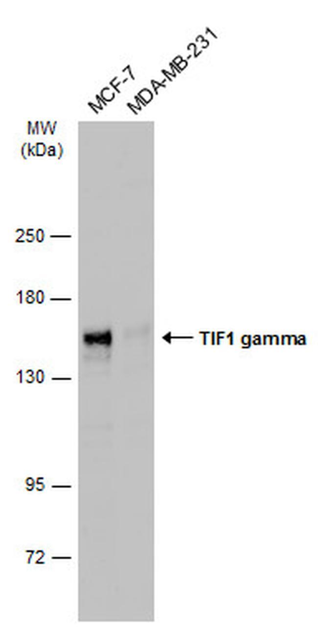
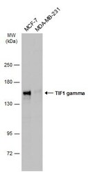
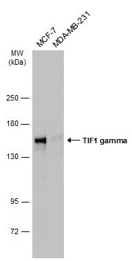
- Western blot analysis of TIF1 gamma in whole cell lysate using 30 µg of protein. Samples were separated with 5% SDS-PAGE and incubated with TIF1 gamma polyclonal antibody (Product # PA5-78496) using a dilution of 1:1000 followed by HRP-conjugated anti-rabbit IgG.
- Submitted by
- Invitrogen Antibodies (provider)
- Main image

- Experimental details
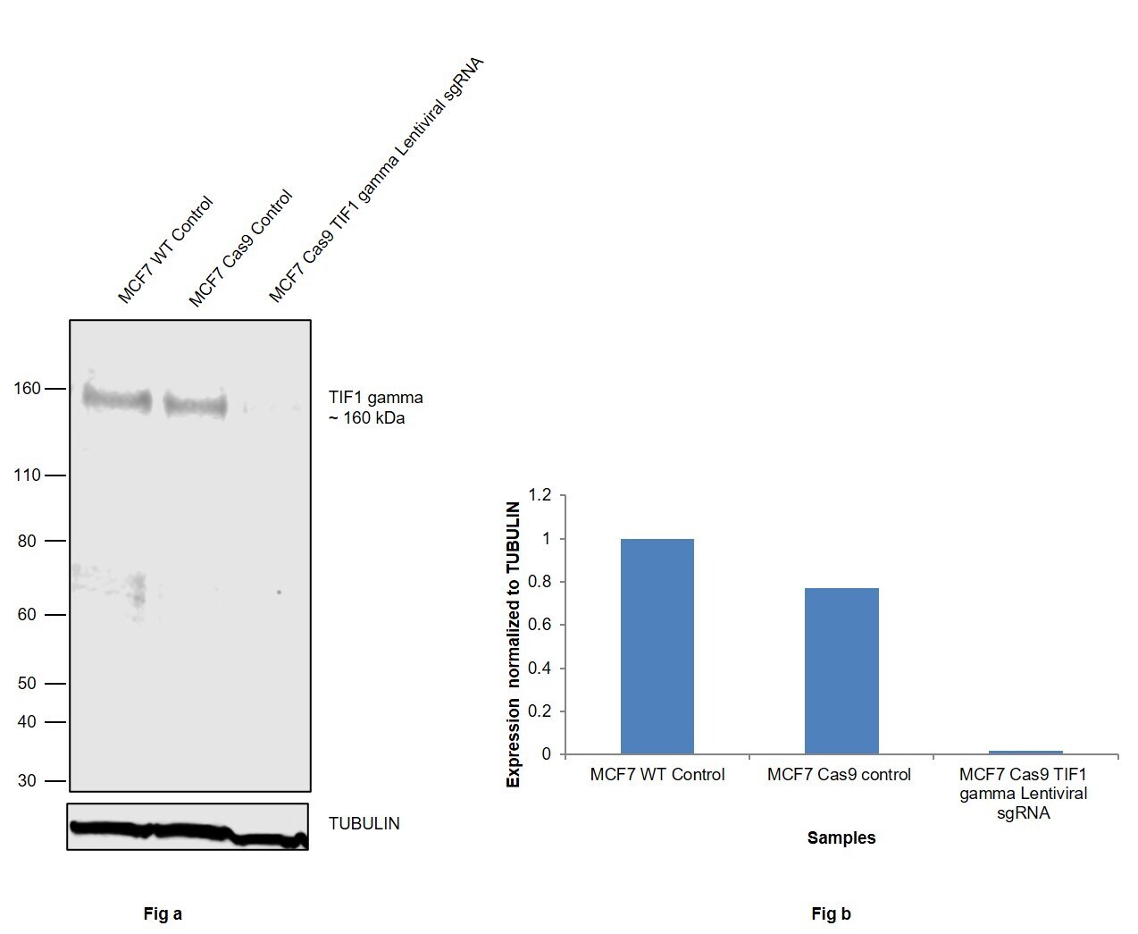
- CRISPR-Cas9 mediated genome editing ofTIF1 gamma (as confirmed by next generation sequencing) was achieved by using LentiArray™ Lentiviral sgRNA (Product # A32042, AssayID CRISPR1029102_LV) and LentiArray Cas9 Lentivirus (Product # A32064). Fig (a) Western blot analysis of TIF1 gamma was performed by loading 30 µg of MCF7 Wild Type (Lane 1), MCF7 Cas9 (Lane 2) and MCF7 Cas9 cells transduced with TIF1 gamma Lentiviral sgRNA (Lane 3) modified whole cell extracts. The samples were electrophoresed using NuPAGE™ 3 to 8%, Tris-Acetate, 1.0 mm, Mini Protein Gel (Product # EC66952BOX). Resolved proteins were then transferred onto a nitrocellulose membrane (Product # IB23001) by iBlot® 2 Dry Blotting System (Product # IB21001). The blot was probed with a TIF1 gamma Polyclonal Antibody (Product # PA5-78496) using 1:1,000 dilution and Goat anti-Rabbit IgG (H+L) Superclonal™ Recombinant Secondary Antibody, HRP (Product # A27036 1:4,000 dilution). Chemiluminescent detection was performed using Novex® ECL Chemiluminescent Substrate Reagent Kit (Product # WP20005). A reduced signal in sgRNA transduced cells using the LentiArray™ CRISPR product line confirms that antibody is specific toTIF1 gamma (Fig (b)).
- Submitted by
- Invitrogen Antibodies (provider)
- Main image
- Experimental details
- Western Blot using TIF1 gamma Polyclonal Antibody (Product # PA5-78496). Various whole cell extracts (30 µg) were separated by 5% SDS-PAGE, and the membrane was blotted with TIF1 gamma Polyclonal Antibody (Product # PA5-78496) diluted at 1:1,000. The HRP-conjugated anti-rabbit IgG antibody was used to detect the primary antibody.
- Submitted by
- Invitrogen Antibodies (provider)
- Main image

- Experimental details
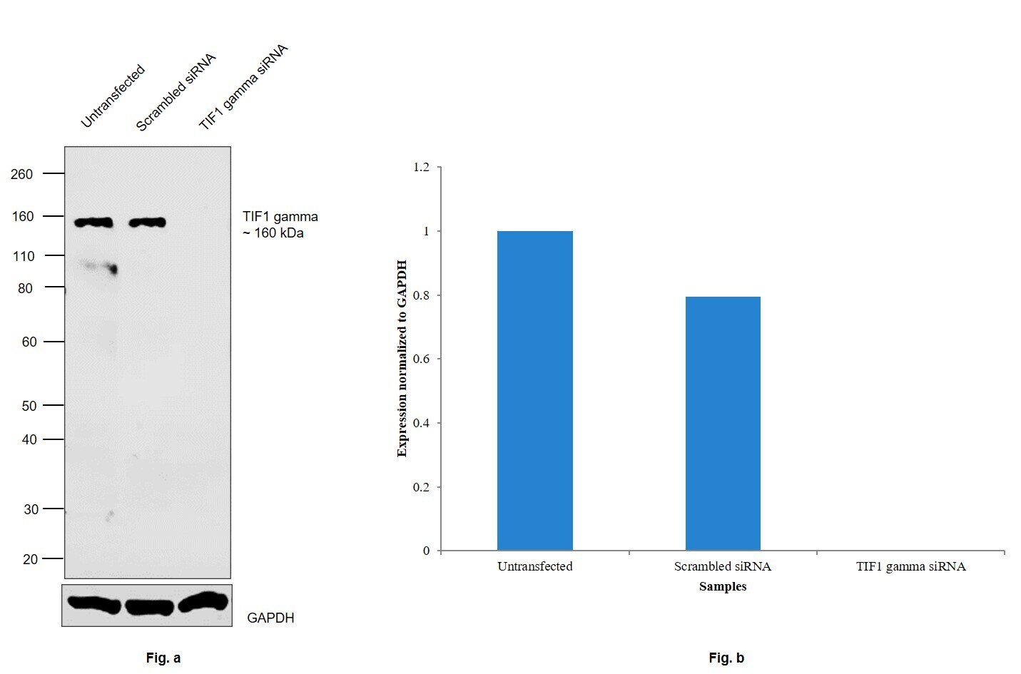
- Knockdown of E3 ubiquitin-protein ligase TRIM33 was achieved by transfecting BeWo with E3 ubiquitin-protein ligase TRIM33 specific siRNAs (Silencer® select Product # S28371, S28372). Western blot analysis (Fig. a) was performed using Modified whole cell extracts (1% SDS) from the E3 ubiquitin-protein ligase TRIM33 knockdown cells (lane 3), non-targeting scrambled siRNA transfected cells (lane 2) and untransfected cells (lane 1). The blot was probed with TIF1 gamma Polyclonal Antibody (Product # PA5-78496, 1:1000 dilution ) and Goat anti-Rabbit IgG (H+L) Superclonal™ Recombinant Secondary Antibody, HRP (Product # A27036, 1:4000 dilution). Densitometric analysis of this western blot is shown in histogram (Fig. b). Loss of signal upon siRNA mediated knock down confirms that antibody is specific to E3 ubiquitin-protein ligase TRIM33.
- Submitted by
- Invitrogen Antibodies (provider)
- Main image

- Experimental details
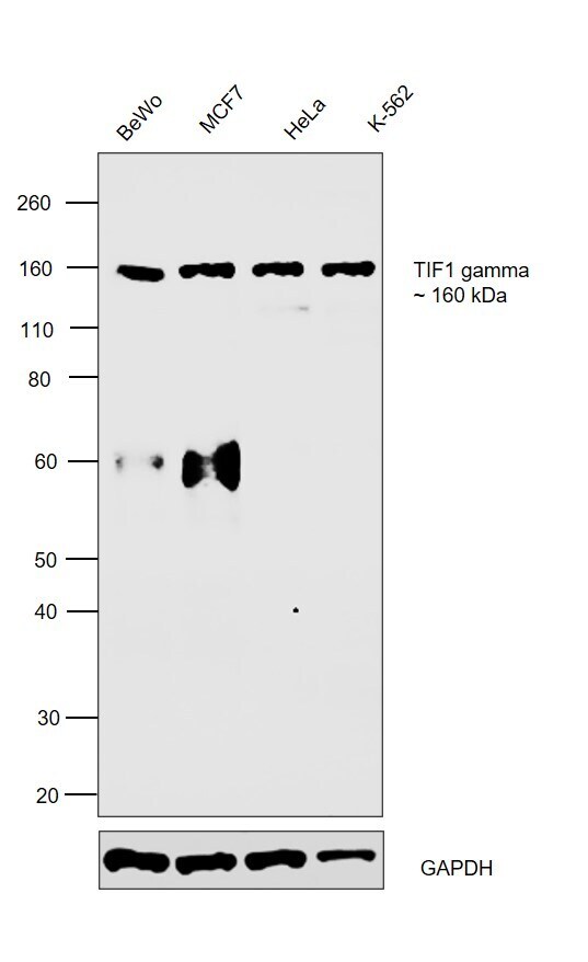
- Western blot was performed using Anti-TIF1 gamma Polyclonal Antibody (Product # PA5-78496) and a 160 kDa band corresponding to E3 ubiquitin-protein ligase TRIM33 was observed across the cell lines tested. Modified whole cell extracts (1% SDS) (30 µg lysate) of BeWo (Lane 1), MCF7 (Lane 2), HeLa (Lane 3) and K-562 (Lane 4) were electrophoresed using NuPAGE™ 4-12% Bis-Tris Protein Gel (Product # NP0321BOX). Resolved proteins were then transferred onto a Nitrocellulose membrane (Product # IB23001) by iBlot® 2 Dry Blotting System (Product # IB21001). The blot was probed with the primary antibody (1:1000 dilution) and detected by chemiluminescence with Goat anti-Rabbit IgG (H+L) Superclonal™ Recombinant Secondary Antibody, HRP (Product # A27036, 1:4000 dilution) using the iBright FL 1000 (Product # A32752). Chemiluminescent detection was performed using Novex® ECL Reagent Kit (Product # WP20005). An uncharacterized band of ~60 kDa was also observed in BeWo and MCF7.
Supportive validation
- Submitted by
- Invitrogen Antibodies (provider)
- Main image

- Experimental details
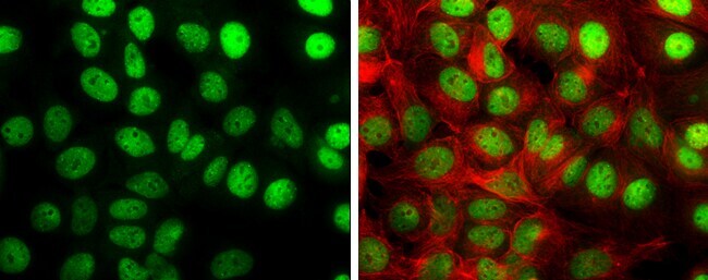
- Immunocytochemistry-Immunofluorescence analysis of TIF1 gamma was performed in MCF 7 cells fixed in 4% paraformaldehyde at RT for 15 min. Green: TIF1 gamma Polyclonal Antibody (Product # PA5 78496) diluted at 1:2000. Red: alpha Tubulin, a cytoskeleton marker.
- Submitted by
- Invitrogen Antibodies (provider)
- Main image

- Experimental details
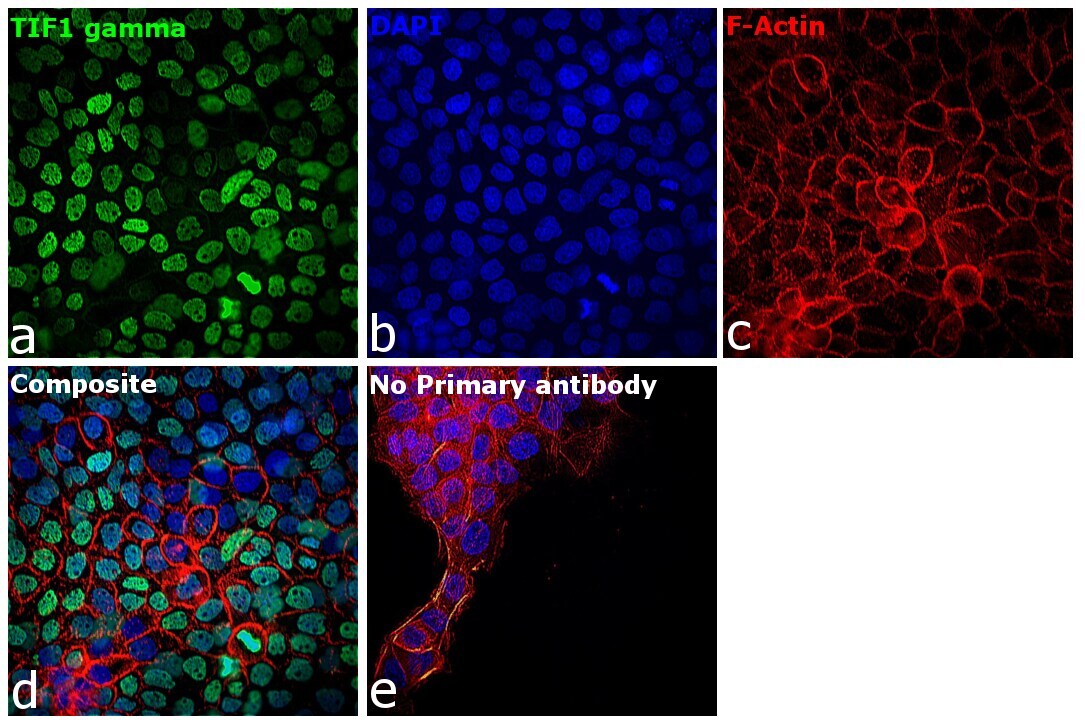
- Immunofluorescence analysis of TIF1 gamma was performed using 70% confluent log phase A-431 cells. The cells were fixed with 4% paraformaldehyde for 10 minutes, permeabilized with 0.1% Triton™ X-100 for 15 minutes, and blocked with 2% BSA for 1 hour at room temperature. The cells were labeled with TIF1 gamma Polyclonal Antibody (Product # PA5-78496) at 1:100 dilution in 0.1% BSA, incubated at 4 degree celsius overnight and then with Donkey anti-Rabbit IgG (H+L) Highly Cross-Adsorbed Secondary Antibody, Alexa Fluor Plus 488 (Product # A32790) at a dilution of 1:2000 for 45 minutes at room temperature (Panel a: green). Nuclei (Panel b: blue) were stained with Hoechst 33342 (Product # H1399). F-actin (Panel c: red) was stained with Rhodamine Phalloidin (Product # R415, 1:300). Panel d represents the merged image showing nuclear localization. Panel e represents control cells with no primary antibody to assess background. The images were captured at 40X magnification in CellInsight CX7 LZR High-Content Screening (HCS) Platform (Product # CX7C1115LZR).
- Submitted by
- Invitrogen Antibodies (provider)
- Main image

- Experimental details
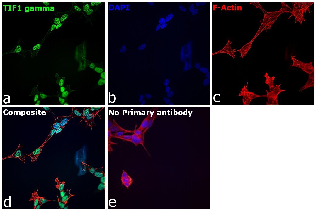
- Immunofluorescence analysis of TIF1 gamma was performed using 70% confluent log phase SH-SY5Y cells. The cells were fixed with 4% paraformaldehyde for 10 minutes, permeabilized with 0.1% Triton™ X-100 for 15 minutes, and blocked with 2% BSA for 1 hour at room temperature. The cells were labeled with TIF1 gamma Polyclonal Antibody (Product # PA5-78496) at 1:100 dilution in 0.1% BSA, incubated at 4 degree celsius overnight and then with Donkey anti-Rabbit IgG (H+L) Highly Cross-Adsorbed Secondary Antibody, Alexa Fluor Plus 488 (Product # A32790) at a dilution of 1:2000 for 45 minutes at room temperature (Panel a: green). Nuclei (Panel b: blue) were stained with Hoechst 33342 (Product # H1399). F-actin (Panel c: red) was stained with Rhodamine Phalloidin (Product # R415, 1:300). Panel d represents the merged image showing nuclear localization. Panel e represents control cells with no primary antibody to assess background. The images were captured at 40X magnification in CellInsight CX7 LZR High-Content Screening (HCS) Platform (Product # CX7C1115LZR).
Supportive validation
- Submitted by
- Invitrogen Antibodies (provider)
- Main image
- Experimental details
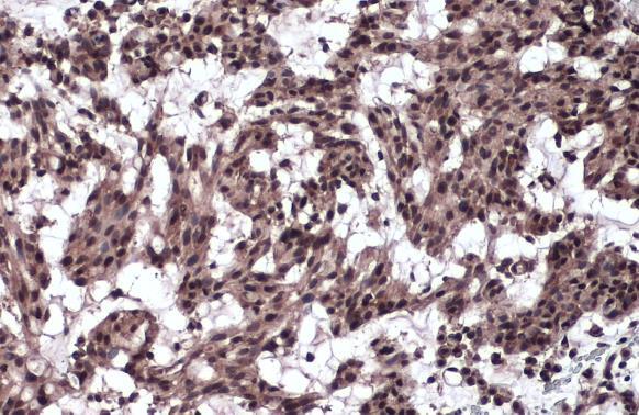
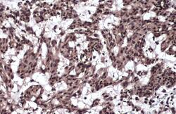
- TIF1 gamma Polyclonal Antibody detects TIF1 gamma protein at nucleus by immunohistochemical analysis. Sample: Paraffin-embedded human breast carcinoma. TIF1 gamma stained by TIF1 gamma Polyclonal Antibody (Product # PA5-78496) diluted at 1:1,000. Antigen Retrieval: Citrate buffer, pH 6.0, 15 min.