Explore
 Validate
 Learn
Learn Western blot
Antibody data
- Antibody Data
- Antigen structure
- References [2]
- Comments [0]
- Validations
- Western blot [1]
- Immunocytochemistry [1]
- Chromatin Immunoprecipitation [1]
Submit
Validation data
Reference
Comment
Report error
- Product number
- AF7119 - Provider product page

- Provider
- R&D Systems
- Product name
- Human SREBP2 Antibody
- Antibody type
- Polyclonal
- Description
- Immunogen affinity purified. Detects human SREBP2 in direct ELISAs and Western blots. In direct ELISAs, less than 1% cross-reactivity with recombinant human (rh) SREBP1B and rhSREBP1C is observed.
- Reactivity
- Human
- Host
- Goat
- Conjugate
- Unconjugated
- Antigen sequence
Q12772- Isotype
- IgG
- Vial size
- 100 ug
- Concentration
- LYOPH
- Storage
- Use a manual defrost freezer and avoid repeated freeze-thaw cycles. 12 months from date of receipt, -20 to -70 °C as supplied. 1 month, 2 to 8 °C under sterile conditions after reconstitution. 6 months, -20 to -70 °C under sterile conditions after reconstitution.
Submitted references The ER membrane protein complex promotes biogenesis of sterol-related enzymes maintaining cholesterol homeostasis.
CAT-2003: A novel sterol regulatory element-binding protein inhibitor that reduces steatohepatitis, plasma lipids, and atherosclerosis in apolipoprotein E*3-Leiden mice.
Volkmar N, Thezenas ML, Louie SM, Juszkiewicz S, Nomura DK, Hegde RS, Kessler BM, Christianson JC
Journal of cell science 2019 Jan 16;132(2)
Journal of cell science 2019 Jan 16;132(2)
CAT-2003: A novel sterol regulatory element-binding protein inhibitor that reduces steatohepatitis, plasma lipids, and atherosclerosis in apolipoprotein E*3-Leiden mice.
Zimmer M, Bista P, Benson EL, Lee DY, Liu F, Picarella D, Vega RB, Vu CB, Yeager M, Ding M, Liang G, Horton JD, Kleemann R, Kooistra T, Morrison MC, Wielinga PY, Milne JC, Jirousek MR, Nichols AJ
Hepatology communications 2017 Jun;1(4):311-325
Hepatology communications 2017 Jun;1(4):311-325
No comments: Submit comment
Supportive validation
- Submitted by
- R&D Systems (provider)
- Main image
- Experimental details
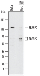
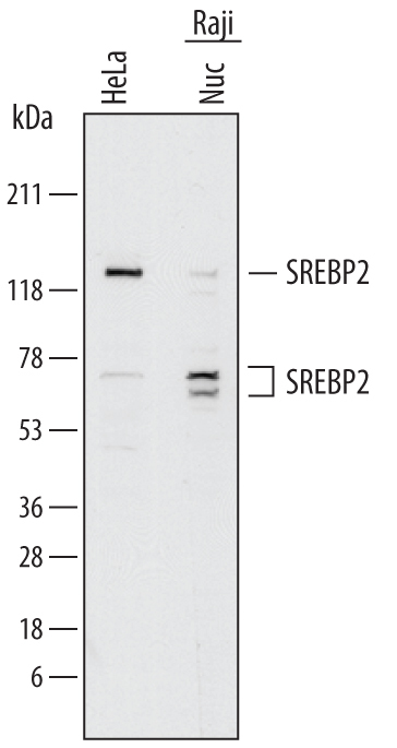
- Detection of Human SREBP2 by Western Blot. Western blot shows lysates of HeLa human cervical epithelial carcinoma cell line and Raji human Burkitt's lymphoma cell line. Gels were loaded with 15 µg of nuclear extracts (Nuc). PVDF membrane was probed with 2 µg/mL of Goat Anti-Human SREBP2 Antigen Affinity-purified Polyclonal Antibody (Catalog # AF7119) followed by HRP-conjugated Anti-Goat IgG Secondary Antibody (Catalog # HAF017). Specific bands were detected for SREBP2 at approximately 125 kDa and 60-70 kDa (as indicated). This experiment was conducted under reducing conditions and using Immunoblot Buffer Group 1.
Supportive validation
- Submitted by
- R&D Systems (provider)
- Main image

- Experimental details
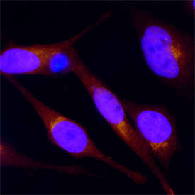
- SREBP2 in HeLa Human Cell Line. SREBP2 was detected in immersion fixed HeLa human cervical epithelial carcinoma cell line using Goat Anti-Human SREBP2 Antigen Affinity-purified Polyclonal Antibody (Catalog # AF7119) at 15 µg/mL for 3 hours at room temperature. Cells were stained using the NorthernLights™ 557-conjugated Anti-Goat IgG Secondary Antibody (red; Catalog # NL001) and counterstained with DAPI (blue). Specific staining was localized to cytoplasm. View our protocol for Fluorescent ICC Staining of Cells on Coverslips.
Supportive validation
- Submitted by
- R&D Systems (provider)
- Main image

- Experimental details
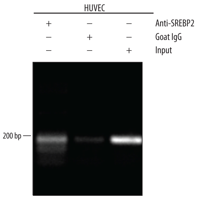
- Detection of SREBP2-regulated Genes by Chromatin Immunoprecipitation. HUVEC human umbilical vein endothelial cells were serum-starved for 5 hours, fixed using formaldehyde, resuspended in lysis buffer, and sonicated to shear chromatin. SREBP2/DNA complexes were immuno-precipitated using 5 μg Goat Anti-Human SREBP2 Antigen Affinity-purified Polyclonal Antibody (Catalog # AF7119) or control antibody (Catalog # AB-108-C) for 15 minutes in an ultrasonic bath, followed by Biotinylated Anti-Goat IgG Secondary Antibody (Catalog # BAF109). Immuno-complexes were captured using 50 μL of MagCellect Streptavidin Ferrofluid (Catalog # MAG999) and DNA was purified using chelating resin solution. The ABCA1 promoter was detected by standard PCR. In the figure, "Input" represents total cell lysate DNA.