Explore
 Validate
 Learn
Learn Western blot
 Immunohistochemistry
Antibody data
- Antibody Data
- Antigen structure
- References [1]
- Comments [0]
- Validations
- Immunohistochemistry [1]
Submit
Validation data
Reference
Comment
Report error
- Product number
- HPA043285 - Provider product page

- Provider
- Atlas Antibodies
- Proper citation
- Atlas Antibodies Cat#HPA043285, RRID:AB_10962482
- Product name
- Anti-HSPA1L
- Antibody type
- Polyclonal
- Description
- Polyclonal Antibody against Human HSPA1L, Gene description: heat shock 70kDa protein 1-like, Alternative Gene Names: HSP70-HOM, hum70t, Validated applications: IHC, WB, Uniprot ID: P34931, Storage: Store at +4°C for short term storage. Long time storage is recommended at -20°C.
- Reactivity
- Human, Mouse, Rat
- Host
- Rabbit
- Conjugate
- Unconjugated
- Isotype
- IgG
- Vial size
- 100 µl
- Concentration
- 0.3 mg/ml
- Storage
- Store at +4°C for short term storage. Long time storage is recommended at -20°C.
- Handling
- The antibody solution should be gently mixed before use.
Submitted references Profiling the Hsp70 Chaperone Network in Heat-Induced Proteotoxic Stress Models of Human Neurons
Alharbi B, Albinhassan T, Alzahrani R, Bouchama A, Mohammad S, Alomari A, Bin-Jumah M, AlSuhaibani E, Malik S
Biology 2023;12(3):416
Biology 2023;12(3):416
No comments: Submit comment
Supportive validation
- Submitted by
- Atlas Antibodies (provider)
- Enhanced method
- Orthogonal validation
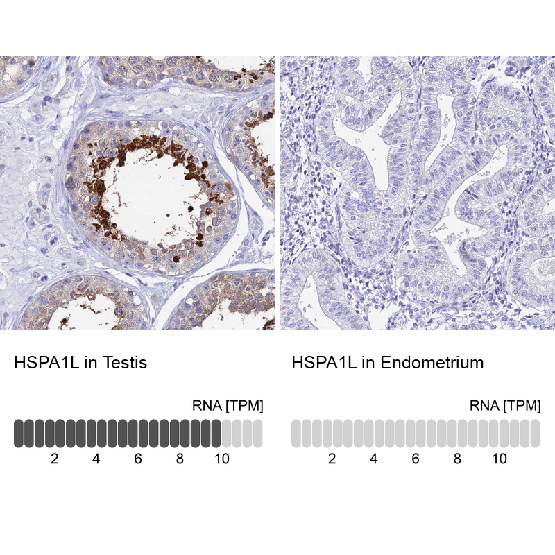
- Main image
- Experimental details
- Immunohistochemistry analysis in human testis and endometrium tissues using HPA043285 antibody. Corresponding HSPA1L RNA-seq data are presented for the same tissues.
- Sample type
- Human
- Protocol
- Protocol