Explore
 Validate
 Learn
Learn Western blot
 Immunocytochemistry
Antibody data
- Antibody Data
- Antigen structure
- References [1]
- Comments [0]
- Validations
- Immunocytochemistry [3]
- Immunohistochemistry [1]
- Other assay [1]
Submit
Validation data
Reference
Comment
Report error
- Product number
- PA5-21939 - Provider product page

- Provider
- Invitrogen Antibodies
- Product name
- Activin A Polyclonal Antibody
- Antibody type
- Polyclonal
- Antigen
- Synthetic peptide
- Description
- Recommended positive controls: A549, H1299, HCT-116. Predicted reactivity: Mouse (100%), Rat (100%), Cat (100%), Pig (100%), Rabbit (100%), Chicken (100%), Sheep (100%), Bovine (100%). Store product as a concentrated solution. Centrifuge briefly prior to opening the vial.
- Reactivity
- Human
- Host
- Rabbit
- Isotype
- IgG
- Vial size
- 100 μL
- Concentration
- 0.66 mg/mL
- Storage
- Store at 4°C short term. For long term storage, store at -20°C, avoiding freeze/thaw cycles.
Submitted references Senescence marker activin A is increased in human diabetic kidney disease: association with kidney function and potential implications for therapy.
Bian X, Griffin TP, Zhu X, Islam MN, Conley SM, Eirin A, Tang H, O'Shea PM, Palmer AK, McCoy RG, Herrmann SM, Mehta RA, Woollard JR, Rule AD, Kirkland JL, Tchkonia T, Textor SC, Griffin MD, Lerman LO, Hickson LJ
BMJ open diabetes research & care 2019;7(1):e000720
BMJ open diabetes research & care 2019;7(1):e000720
No comments: Submit comment
Supportive validation
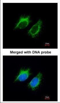
- Submitted by
- Invitrogen Antibodies (provider)
- Main image
- Experimental details
- Immunofluorescent analysis of Inhibin beta-A in paraformaldehyde-fixed HeLa cells using an Inhibin beta-A polyclonal antibody (Product # PA5-21939) at a 1:200 dilution.

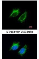
- Submitted by
- Invitrogen Antibodies (provider)
- Main image

- Experimental details
- Immunofluorescence analysis of paraformaldehyde-fixed HeLa, using Inhibin beta-A (Product # PA5-21939) antibody at 1:200 dilution.
- Submitted by
- Invitrogen Antibodies (provider)
- Main image

- Experimental details
- Immunofluorescence analysis of paraformaldehyde-fixed HeLa, using Inhibin beta-A (Product # PA5-21939) antibody at 1:200 dilution.

Supportive validation
- Submitted by
- Invitrogen Antibodies (provider)
- Main image
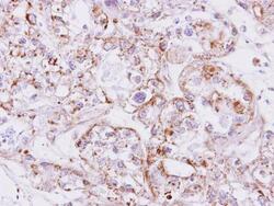
- Experimental details
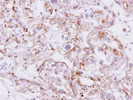
- Immunohistochemical analysis of paraffin-embedded CLEAR CELL OVCA xenograft, using Inhibin beta A (Product # PA5-21939) antibody at 1:500 dilution. Antigen Retrieval: EDTA based buffer, pH 8.0, 15 min.
Supportive validation
- Submitted by
- Invitrogen Antibodies (provider)
- Main image
- Experimental details
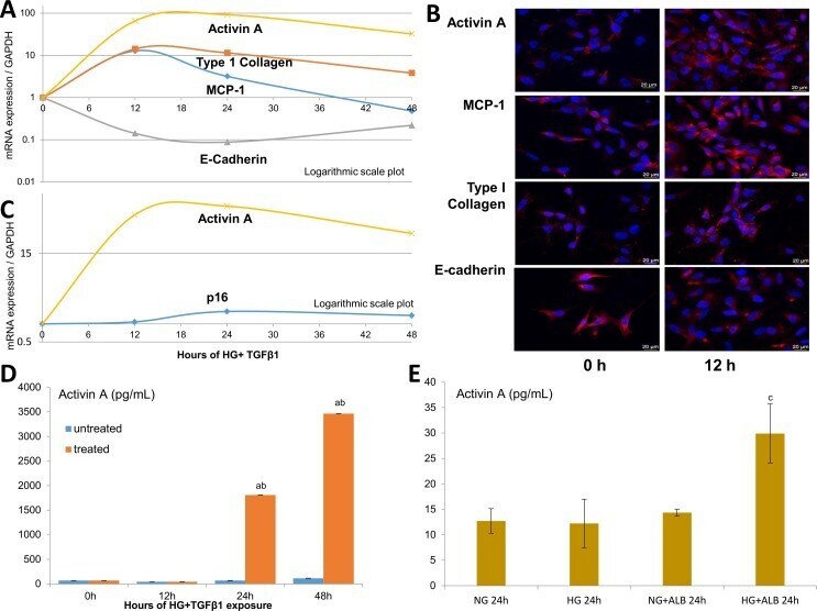
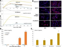
- Figure 5 In vitro models of DKD-related injury. HK-2 studies following incubation with high glucose (HG, 25 mmol/L), transforming growth factor-beta1 (TGF-beta1; 5 ng/mL) and albumin (ALB, 5 mg/mL). Time-dependent effect of HG+TGF-beta1 on the mRNA/gene expression of activin A, monocyte chemoattractant protein-1 (MCP-1; an inflammatory marker), type I collagen (mesenchymal marker) and E-cadherin (epithelial marker) in injured HK-2 cells (A) Immunofluorescent staining of HK-2 cells injured by HG+TGF-beta1 at 12 hours (h) compared with baseline (B) Time-dependent effect for activin A and p16 expression in injured HK-2 cells (C) Protein levels of activin A in the supernatant of HK-2 treated with HG+TGF-beta1 (D) and combinations of HG with or without ALB (E) at 24 hours. Values are mean+-SEM. a P