Explore
 Validate
 Learn
Learn Western blot
 ELISA
ELISA Immunocytochemistry
Antibody data
- Antibody Data
- Antigen structure
- References [0]
- Comments [0]
- Validations
- Immunocytochemistry [2]
- Immunohistochemistry [3]
Submit
Validation data
Reference
Comment
Report error
- Product number
- PA5-90073 - Provider product page

- Provider
- Invitrogen Antibodies
- Product name
- G3BP1 Polyclonal Antibody
- Antibody type
- Polyclonal
- Antigen
- Recombinant full-length protein
- Description
- Immunogen sequence: FRYQDEVFGG FVTEPQEESE EEVEEPEERQ QTPEVVPDDS GTFYDQAVVS NDMEEHLEEP VAEPEPDPEP EPEQEPVSEI QEEKPEPVLE ETAPEDAQKS SSPAPADIAQ TVQEDLRTFS WASVTSKNLP PSGAVPVTGI PPHVVKVPAS QPRPESKPES QIPPQRPQRD QRVREQRINI PPQRGPRPIR EAGEQGDIEP; Positive Samples: A-431, HeLa, 293T, Jurka, MCF7; Cellular Location: Cell membrane, Cytoplasm, Cytoplasmic granule, Nucleus, cytosol
- Reactivity
- Human, Mouse, Rat
- Host
- Rabbit
- Isotype
- IgG
- Vial size
- 100 μL
- Concentration
- 1.75 mg/mL
- Storage
- -20°C, Avoid Freeze/Thaw Cycles
No comments: Submit comment
Supportive validation
- Submitted by
- Invitrogen Antibodies (provider)
- Main image

- Experimental details
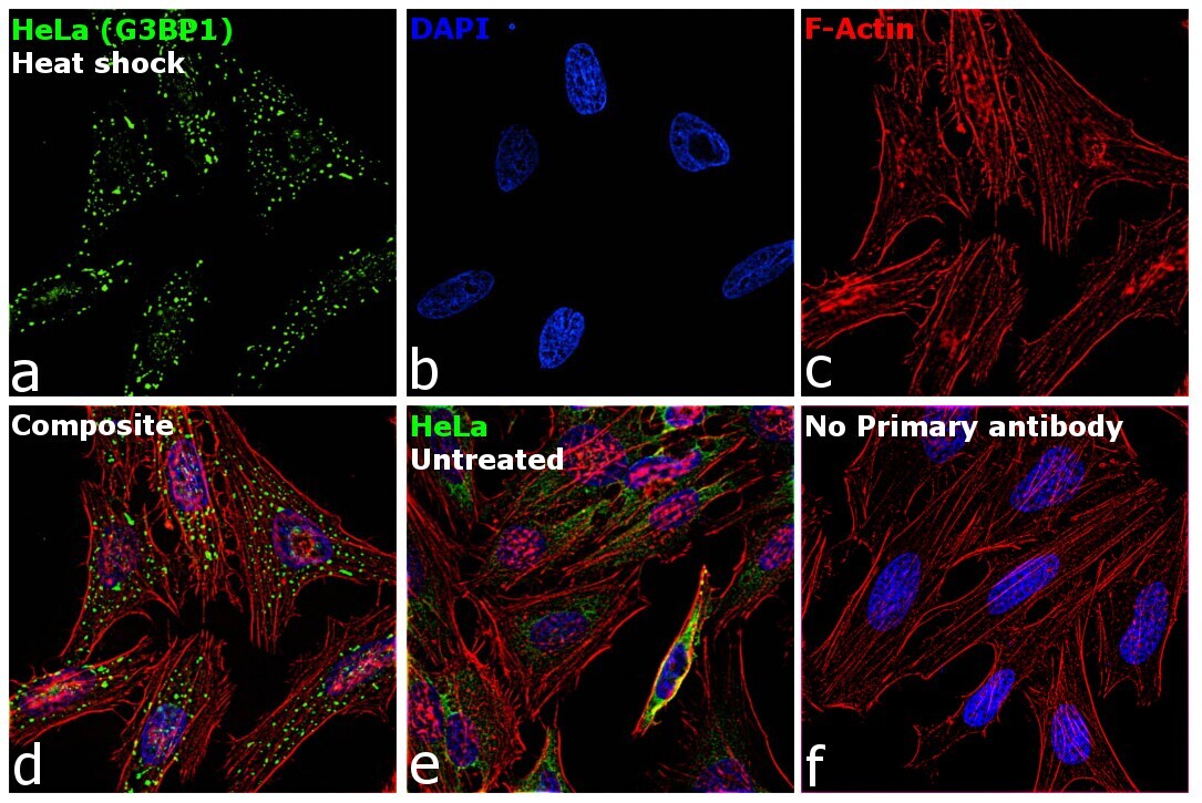
- Immunofluorescence analysis of Ras GTPase-activating protein-binding protein 1 was performed using 70% confluent log phase HeLa cells treated with heat shock at 45 degree centigrade for 60 minutes. The cells were fixed with 4% paraformaldehyde for 5 minutes, permeabilized with 0.1% Triton™ X-100 for 10 minutes, and blocked with 2% BSA for overnight at room temperature. The cells were labeled with G3BP1 Polyclonal Antibody (Product # PA5-90073) at 1:100 dilution in 0.1% BSA, incubated at 4 degree celsius overnight and then labeled with Donkey anti-Rabbit IgG (H+L) Highly Cross-Adsorbed Secondary Antibody, Alexa Fluor Plus 488 (Product # A32790), (1:2000 dilution), for 45 minutes at room temperature (Panel a: Green). Nuclei (Panel b: Blue) were stained with ProLong™ Diamond Antifade Mountant with DAPI (Product # P36962). F-actin (Panel c: Red) was stained with Rhodamine Phalloidin (Product # R415, 1:300). Panel d represents the merged image showing stress granule localization. Panel e represents control untreated cells showing cytoplasmic localization. Panel f represents control cells with no primary antibody to assess background. The images were captured at 60X magnification.
- Submitted by
- Invitrogen Antibodies (provider)
- Main image
- Experimental details
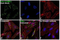
- Immunofluorescence analysis of Ras GTPase-activating protein-binding protein 1 was performed using 70% confluent log phase HeLa cells treated with heat shock at 45 degree centigrade for 60 minutes. The cells were fixed with 4% paraformaldehyde for 5 minutes, permeabilized with 0.1% Triton™ X-100 for 10 minutes, and blocked with 2% BSA for overnight at room temperature. The cells were labeled with G3BP1 Polyclonal Antibody (Product # PA5-90073) at 1:100 dilution in 0.1% BSA, incubated at 4 degree celsius overnight and then labeled with Donkey anti-Rabbit IgG (H+L) Highly Cross-Adsorbed Secondary Antibody, Alexa Fluor Plus 488 (Product # A32790), (1:2000 dilution), for 45 minutes at room temperature (Panel a: Green). Nuclei (Panel b: Blue) were stained with ProLong™ Diamond Antifade Mountant with DAPI (Product # P36962). F-actin (Panel c: Red) was stained with Rhodamine Phalloidin (Product # R415, 1:300). Panel d represents the merged image showing stress granule localization. Panel e represents control untreated cells showing cytoplasmic localization. Panel f represents control cells with no primary antibody to assess background. The images were captured at 60X magnification.

Supportive validation
- Submitted by
- Invitrogen Antibodies (provider)
- Main image
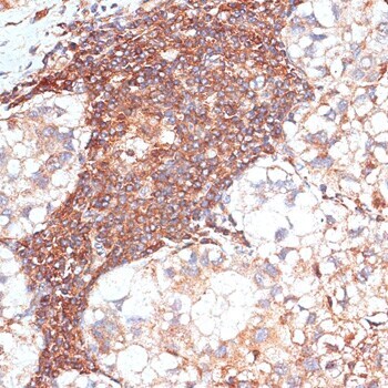
- Experimental details
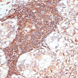
- Immunohistochemistry analysis of G3BP1 in paraffin-embedded human liver cancer. Samples were incubated with G3BP1 Polyclonal antibody (Product # PA5-90073) using a dilution of 1:100 (40x lens). Perform microwave antigen retrieval with 10 mM PBS buffer pH 7.2 before commencing with IHC staining protocol.
- Submitted by
- Invitrogen Antibodies (provider)
- Main image

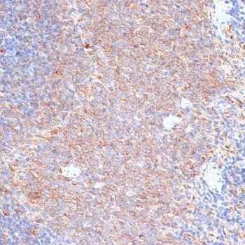
- Experimental details
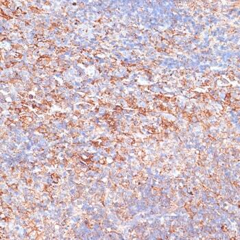
- Immunohistochemistry analysis of G3BP1 in paraffin-embedded rat spleen. Samples were incubated with G3BP1 Polyclonal antibody (Product # PA5-90073) using a dilution of 1:100 (40x lens). Perform microwave antigen retrieval with 10 mM PBS buffer pH 7.2 before commencing with IHC staining protocol.
- Submitted by
- Invitrogen Antibodies (provider)
- Main image
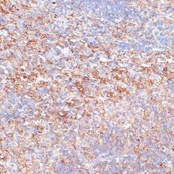
- Experimental details
- Immunohistochemistry analysis of G3BP1 in paraffin-embedded mouse spleen. Samples were incubated with G3BP1 Polyclonal antibody (Product # PA5-90073) using a dilution of 1:100 (40x lens). Perform microwave antigen retrieval with 10 mM PBS buffer pH 7.2 before commencing with IHC staining protocol.