Explore
 Validate
 Learn
Learn Western blot
 Immunocytochemistry
Antibody data
- Antibody Data
- Antigen structure
- References [6]
- Comments [0]
- Validations
- Immunocytochemistry [5]
- Immunohistochemistry [2]
- Other assay [3]
Submit
Validation data
Reference
Comment
Report error
- Product number
- PA5-29455 - Provider product page

- Provider
- Invitrogen Antibodies
- Product name
- G3BP1 Polyclonal Antibody
- Antibody type
- Polyclonal
- Antigen
- Recombinant full-length protein
- Description
- Recommended positive controls: 293T, A431, HeLa, HepG2. Predicted reactivity: Mouse (90%), Rat (91%), Pig (94%), Chicken (82%), Rhesus Monkey (99%), Bovine (95%). Store product as a concentrated solution. Centrifuge briefly prior to opening the vial.
- Reactivity
- Human, Mouse, Rat
- Host
- Rabbit
- Isotype
- IgG
- Vial size
- 100 μL
- Concentration
- 0.35 mg/mL
- Storage
- Store at 4°C short term. For long term storage, store at -20°C, avoiding freeze/thaw cycles.
Submitted references Identification of a novel compound that simultaneously impairs the ubiquitin-proteasome system and autophagy.
Small-molecule modulators of INAVA cytosolic condensate and cell-cell junction assemblies.
TDP-43, a protein central to amyotrophic lateral sclerosis, is destabilized by tankyrase-1 and -2.
Human Insulin Growth Factor 2 mRNA Binding Protein 2 Increases MicroRNA 33a/b Inhibition of Liver ABCA1 Expression and Alters Low-Density Apolipoprotein Levels in Mice.
RNA-Induced Conformational Switching and Clustering of G3BP Drive Stress Granule Assembly by Condensation.
An aberrant phase transition of stress granules triggered by misfolded protein and prevented by chaperone function.
Giovannucci TA, Salomons FA, Stoy H, Herzog LK, Xu S, Qian W, Merino LG, Gierisch ME, Haraldsson M, Lystad AH, Uvell H, Simonsen A, Gustavsson AL, Vallin M, Dantuma NP
Autophagy 2022 Jul;18(7):1486-1502
Autophagy 2022 Jul;18(7):1486-1502
Small-molecule modulators of INAVA cytosolic condensate and cell-cell junction assemblies.
Chang D, Luong P, Li Q, LeBarron J, Anderson M, Barrett L, Lencer WI
The Journal of cell biology 2021 Sep 6;220(9)
The Journal of cell biology 2021 Sep 6;220(9)
TDP-43, a protein central to amyotrophic lateral sclerosis, is destabilized by tankyrase-1 and -2.
McGurk L, Rifai OM, Bonini NM
Journal of cell science 2020 Jun 23;133(12)
Journal of cell science 2020 Jun 23;133(12)
Human Insulin Growth Factor 2 mRNA Binding Protein 2 Increases MicroRNA 33a/b Inhibition of Liver ABCA1 Expression and Alters Low-Density Apolipoprotein Levels in Mice.
Yang M, Gallo-Ebert C, Hayward M, Liu W, McDonough V, Nickels JT Jr
Molecular and cellular biology 2020 Jul 29;40(16)
Molecular and cellular biology 2020 Jul 29;40(16)
RNA-Induced Conformational Switching and Clustering of G3BP Drive Stress Granule Assembly by Condensation.
Guillén-Boixet J, Kopach A, Holehouse AS, Wittmann S, Jahnel M, Schlüßler R, Kim K, Trussina IREA, Wang J, Mateju D, Poser I, Maharana S, Ruer-Gruß M, Richter D, Zhang X, Chang YT, Guck J, Honigmann A, Mahamid J, Hyman AA, Pappu RV, Alberti S, Franzmann TM
Cell 2020 Apr 16;181(2):346-361.e17
Cell 2020 Apr 16;181(2):346-361.e17
An aberrant phase transition of stress granules triggered by misfolded protein and prevented by chaperone function.
Mateju D, Franzmann TM, Patel A, Kopach A, Boczek EE, Maharana S, Lee HO, Carra S, Hyman AA, Alberti S
The EMBO journal 2017 Jun 14;36(12):1669-1687
The EMBO journal 2017 Jun 14;36(12):1669-1687
No comments: Submit comment
Supportive validation
- Submitted by
- Invitrogen Antibodies (provider)
- Main image
- Experimental details
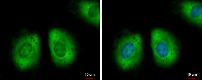
- Immunofluorescent analysis of G3BP1 showing staining in the cytoplasm of HeLa cells. HeLa cells were fixed in 4% paraformaldehyde at RT for 15 min and stained using a G3BP1 polyclonal antibody (Product # PA5-29455) diluted at 1:500. Blue: Hoechst 33342 staining.
- Submitted by
- Invitrogen Antibodies (provider)
- Main image
- Experimental details
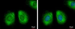
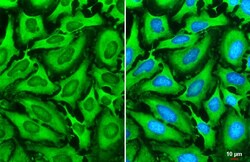
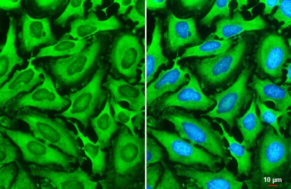
- G3BP1 Polyclonal Antibody detects G3BP1 protein at cytoplasm by immunofluorescent analysis. Sample: HeLa cells were fixed in 4% paraformaldehyde at RT for 15 min. Green: G3BP1 stained by G3BP1 Polyclonal Antibody (Product # PA5-29455) diluted at 1:2,000. Blue: Fluoroshield with DAPI . Scale bar= 10 µm.

- Submitted by
- Invitrogen Antibodies (provider)
- Main image

- Experimental details
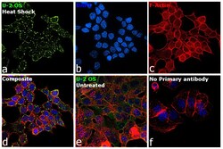
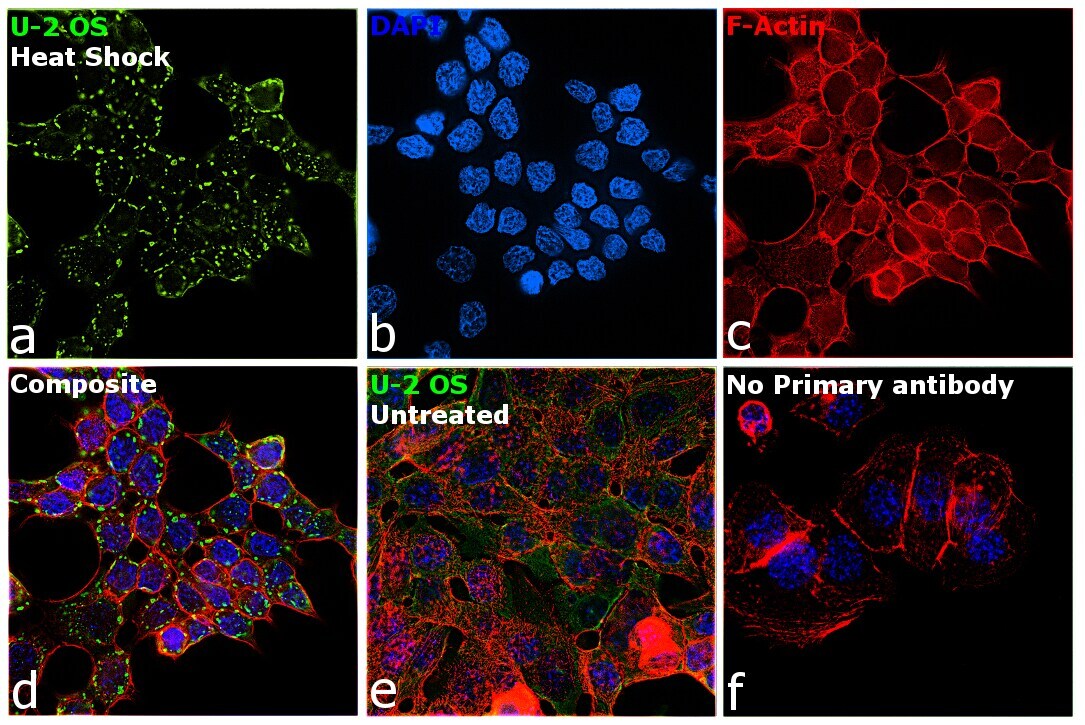
- Immunofluorescence analysis of G3BP1 was performed using 70% confluent log phase U-2 OS treated with Heat Shock (45 degree celsius, 45 minutes). The cells were fixed with 4% paraformaldehyde for 10 minutes, permeabilized with 0.1% Triton™ X-100 for 15 minutes, and blocked with 2% BSA for 45 minutes at room temperature. The cells were labeled with G3BP1 Polyclonal Antibody (Product # PA5-29455) at 1:200 dilution in 0.1% BSA, incubated at 4 degree celsius overnight and then labeled with Donkey anti-Rabbit IgG (H+L) Highly Cross-Adsorbed Secondary Antibody, Alexa Fluor Plus 488 (Product # A32790), (1:2000 dilution), for 45 minutes at room temperature (Panel a: Green). Nuclei (Panel b:Blue) were stained with ProLong™ Diamond Antifade Mountant with DAPI (Product # P36962). F-actin (Panel c: Red) was stained with Rhodamine Phalloidin (Product # R415, 1:300). Panel d represents the merged image showing localization of G3BP1 to stress granules. Panel e represents untreated cells exhibiting cytoplasmic localization. Panel f represents control cells with no primary antibody to assess background. The images were captured at 60x magnification.

- Submitted by
- Invitrogen Antibodies (provider)
- Main image

- Experimental details
- G3BP1 Polyclonal Antibody detects G3BP1 protein at cytoplasm by immunofluorescent analysis. Sample: HeLa cells were fixed in 4% paraformaldehyde at RT for 15 min. Green: G3BP1 stained by G3BP1 Polyclonal Antibody (Product # PA5-29455) diluted at 1:2,000. Blue: Fluoroshield with DAPI . Scale bar= 10 µm.
- Submitted by
- Invitrogen Antibodies (provider)
- Main image
- Experimental details
- Immunofluorescence analysis of G3BP1 was performed using 70% confluent log phase U-2 OS treated with Heat Shock (45 degree celsius, 45 minutes). The cells were fixed with 4% paraformaldehyde for 10 minutes, permeabilized with 0.1% Triton™ X-100 for 15 minutes, and blocked with 2% BSA for 45 minutes at room temperature. The cells were labeled with G3BP1 Polyclonal Antibody (Product # PA5-29455) at 1:200 dilution in 0.1% BSA, incubated at 4 degree celsius overnight and then labeled with Donkey anti-Rabbit IgG (H+L) Highly Cross-Adsorbed Secondary Antibody, Alexa Fluor Plus 488 (Product # A32790), (1:2000 dilution), for 45 minutes at room temperature (Panel a: Green). Nuclei (Panel b:Blue) were stained with ProLong™ Diamond Antifade Mountant with DAPI (Product # P36962). F-actin (Panel c: Red) was stained with Rhodamine Phalloidin (Product # R415, 1:300). Panel d represents the merged image showing localization of G3BP1 to stress granules. Panel e represents untreated cells exhibiting cytoplasmic localization. Panel f represents control cells with no primary antibody to assess background. The images were captured at 60x magnification.
Supportive validation
- Submitted by
- Invitrogen Antibodies (provider)
- Main image
- Experimental details
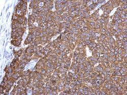
- Immunohistochemical analysis of paraffin-embedded Hela xenograft, using G3BP1 (Product # PA5-29455) antibody at 1:500 dilution. Antigen Retrieval: EDTA based buffer, pH 8.0, 15 min.

- Submitted by
- Invitrogen Antibodies (provider)
- Main image

- Experimental details
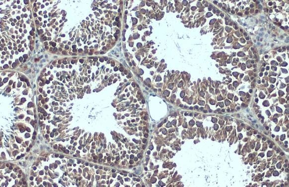
- G3BP1 Polyclonal Antibody detects G3BP1 protein at cytoplasm by immunohistochemical analysis. Sample: Paraffin-embedded mouse testis. G3BP1 stained by G3BP1 Polyclonal Antibody (Product # PA5-29455) diluted at 1:500. Antigen Retrieval: Citrate buffer, pH 6.0, 15 min.
Supportive validation
- Submitted by
- Invitrogen Antibodies (provider)
- Main image

- Experimental details
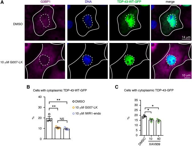
- Fig. 7. Tnks-1/2 inhibition promotes nuclear localization of TDP-43. (A) Treatment with Tnks-1/2 inhibitor G007-LK (10 uM) reduced cytoplasmic accumulation of TDP-43-WT-GFP. Cells were immunostained for the cytoplasmic protein G3BP1 (magenta) and counterstained with Hoechst 33342 (blue). Inner dashed line marks the nuclear boundary and outer solid line marks the cytoplasmic boundary. (B) Treatment of cells with Tnks-1/2 inhibitors G007-LK and IWR1-endo led to a significant reduction in the percentage of cells with cytoplasmic TDP-43-GFP. The percentage of cells with cytoplasmic TDP-43-GFP showing a diffuse GFP signal or GFP-positive puncta was quantified. Mean+-s.e.m. of three independent experiments. One-way ANOVA ( P =0.0032) and a Tukey's tests. (C) Treatment with the Tnks-1/2 inhibitor XAV939 led to a significant reduction in the percentage of cells with cytoplasmic TDP-43-GFP. Mean+-s.e.m. of three independent experiments. One-way ANOVA ( P
- Submitted by
- Invitrogen Antibodies (provider)
- Main image

- Experimental details
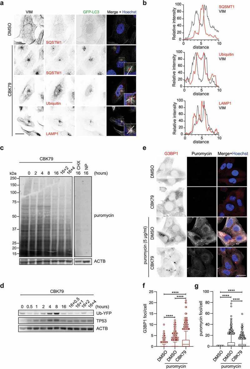
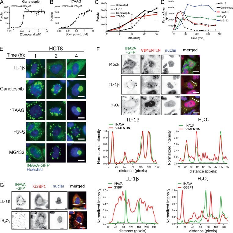
- Figure 3. ROS and HSP90 and proteasome inhibitors induce INAVA puncta. (A and B) Dose response for HSP90 inhibitors (A) ganetespib and (B) 17AAG, mean +- SEM, n = 3. (C) Time course of puncta formation by IL-1beta (10 ng/ml), ganetespib (1 muM) and 17AAG (10 muM), mean +- SD, n = 2. (D) Time course of puncta formation and resolution after treatment with agonists noted above, n = 2. (E) Time course images from D. Scale bar = 10 mum. (F and G) Confocal images of HCT8-INAVA-GFP cells stained for (F) vimentin or (G) G3BP1 following treatment with IL-1beta or H 2 O 2 for 90 min. Hoechst (nuclei). Line scans (arrows) display relative fluorescence intensity of INAVA and (F) vimentin or (G) G3BP1. Scale bar = 20 mum.
- Submitted by
- Invitrogen Antibodies (provider)
- Main image
- Experimental details
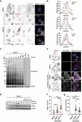
- Figure 5. CBK79 induces proteotoxic stress. (A) Representative images of HOS GFP-LC3B cells treated with DMSO 0.1% or CBK79 (10 uM) for 4 h. Cells were fixed and immunostained using antibodies against VIM, SQSTM1, ubiquitin or LAMP1. Scale bar: 20 um. (B) Line scans at the aggresome sites are shown to visualize the spatial distribution of the indicated proteins (red curves) compared to VIM (gray curves). Intensities are normalized to percentages were 0% = minimum intensity value and 100% = maximum intensity value. (C) MelJuSo Ub-YFP cells were treated with DMSO 0.1% or CBK79 (10 uM) for the indicated timepoints. Two samples were treated for 16 h and then treated with fresh compound solution for the indicated timepoints (""16 h+""). Fifteen minutes before harvesting, puromycin (5 ug/ml) was added to the cells to monitor its incorporation into newly synthesized proteins. Cycloheximide (CHX, 50 ug/ml) was included as control. An untreated sample without puromycin was added (""NP"") as a technical control. Cell lysates were analyzed by immunoblotting with the indicated antibodies. Beta-actin (ACTB) is shown as loading control. Representative blots from one of two independent experiments are shown. (D) MelJuSo Ub-YFP cells were treated with DMSO 0.1% or CBK79 (10 uM) for the indicated timepoints. Two samples were treated for 16 h and then treated with fresh compound solution for the indicated timepoints (""16 h+ re-addition""). Cell lysates were analyzed by immunoblotting with the