 Explore
 Validate
 Learn
Learn Western blot
 Immunocytochemistry
 Immunohistochemistry
Antibody data
- Antibody Data
- Antigen structure
- References [2]
- Comments [0]
- Validations
- Western blot [1]
- Immunocytochemistry [1]
Submit
Validation data
Reference
Comment
Report error
- Product number
- HPA004052 - Provider product page

- Provider
- Atlas Antibodies
- Proper citation
- Atlas Antibodies Cat#HPA004052, RRID:AB_1849348
- Product name
- Anti-G3BP1
- Antibody type
- Polyclonal
- Description
- Polyclonal Antibody against Human G3BP1, Gene description: GTPase activating protein (SH3 domain) binding protein 1, Alternative Gene Names: G3BP, HDH-VIII, Validated applications: ICC, IHC, WB, Uniprot ID: Q13283, Storage: Store at +4°C for short term storage. Long time storage is recommended at -20°C.
- Reactivity
- Human, Mouse, Rat
- Host
- Rabbit
- Conjugate
- Unconjugated
- Isotype
- IgG
- Vial size
- 100 µl
- Concentration
- 0.1 mg/ml
- Storage
- Store at +4°C for short term storage. Long time storage is recommended at -20°C.
- Handling
- The antibody solution should be gently mixed before use.
Submitted references Colocalization of TDP‐43 and stress granules at the early stage of TDP‐43 aggregation in amyotrophic lateral sclerosis
ALS-FTLD-linked mutations of SQSTM1/p62 disrupt selective autophagy and NFE2L2/NRF2 anti-oxidative stress pathway
Mori F, Yasui H, Miki Y, Kon T, Arai A, Kurotaki H, Tomiyama M, Wakabayashi K
Brain Pathology 2023;34(2)
Brain Pathology 2023;34(2)
ALS-FTLD-linked mutations of SQSTM1/p62 disrupt selective autophagy and NFE2L2/NRF2 anti-oxidative stress pathway
Deng Z, Lim J, Wang Q, Purtell K, Wu S, Palomo G, Tan H, Manfredi G, Zhao Y, Peng J, Hu B, Chen S, Yue Z
Autophagy 2019;16(5):917-931
Autophagy 2019;16(5):917-931
No comments: Submit comment
Enhanced validation
- Submitted by
- Atlas Antibodies (provider)
- Enhanced method
- Genetic validation
- Main image

- Experimental details
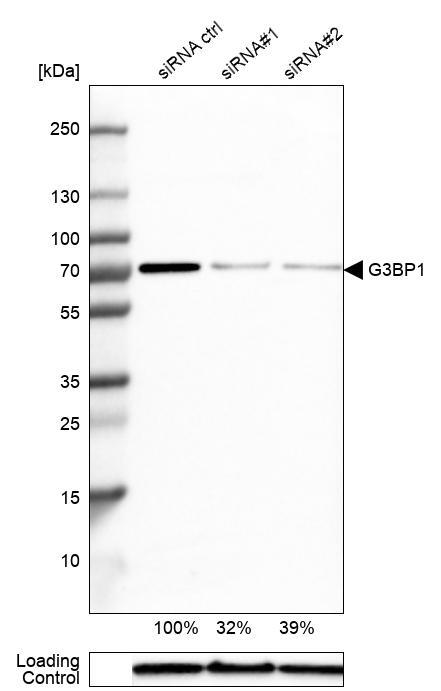
- Western blot analysis in A-431 cells transfected with control siRNA, target specific siRNA probe #1 and #2, using Anti-G3BP1 antibody. Remaining relative intensity is presented. Loading control: Anti-GAPDH.
- Sample type
- Human
- Protocol
- Protocol
Supportive validation
- Submitted by
- Atlas Antibodies (provider)
- Main image
- Experimental details
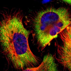
- Immunofluorescent staining of human cell line U-251 MG shows localization to cytosol.
- Sample type
- Human
