Explore
 Validate
 Learn
Learn Western blot
Antibody data
- Antibody Data
- Antigen structure
- References [1]
- Comments [0]
- Validations
- Western blot [2]
- Immunocytochemistry [1]
- Immunoprecipitation [2]
- Immunohistochemistry [2]
Submit
Validation data
Reference
Comment
Report error
- Product number
- GTX101763 - Provider product page

- Provider
- GeneTex
- Proper citation
- GeneTex Cat#GTX101763, RRID:AB_1241420
- Product name
- TUFM antibody
- Antibody type
- Polyclonal
- Reactivity
- Human, Mouse
- Host
- Rabbit
Submitted references Resources for the Comprehensive Discovery of Functional RNA Elements.
Sundararaman B, Zhan L, Blue SM, Stanton R, Elkins K, Olson S, Wei X, Van Nostrand EL, Pratt GA, Huelga SC, Smalec BM, Wang X, Hong EL, Davidson JM, Lécuyer E, Graveley BR, Yeo GW
Molecular cell 2016 Mar 17;61(6):903-13
Molecular cell 2016 Mar 17;61(6):903-13
No comments: Submit comment
Supportive validation
- Submitted by
- GeneTex (provider)
- Main image
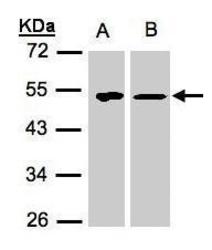
- Experimental details
- Sample(30 ?g of whole cell lysate)A:293TB:H1299 10% SDS PAGEGTX101763 diluted at 1:1500The HRP-conjugated anti-rabbit IgG antibody (GTX213110-01) was used to detect the primary antibody.
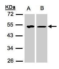
- Submitted by
- GeneTex (provider)
- Main image
- Experimental details
- Sample (30 ?g of whole cell lysate) A:NIH-3T3 10% SDS PAGE GTX101763 diluted at 1:1000 The HRP-conjugated anti-rabbit IgG antibody (GTX213110-01) was used to detect the primary antibody.

Supportive validation
- Submitted by
- GeneTex (provider)
- Main image
- Experimental details
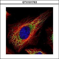
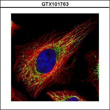
- Confocal immunofluorescence analysis (Olympus FV10i) of paraformaldehyde-fixed HeLa, using TUFM(GTX101763) antibody (Green) at 1:500 dilution. Alpha-tubulin filaments were labeled with GTX11304 (Red) at 1:2500.
Supportive validation
- Submitted by
- GeneTex (provider)
- Main image
- Experimental details
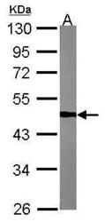
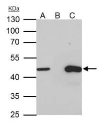
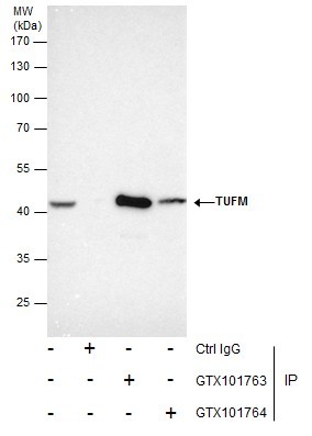
- TUFM antibody immunoprecipitates TUFM protein in IP experiments.IP samples: HepG2 whole cell extractA. 35 £gg HepG2 whole cell extractB. Control with 4 £gg of preimmune Rabbit IgGC. Immunoprecipitation of TUFM protein by 4 £gg TUFM antibody (GTX101763)10 % SDS-PAGEThe immunoprecipitated TUFM protein was detected by TUFM antibody (GTX101763) diluted at 1:1000.[EasyBlot anti-rabbit IgG (GTX221666-01) was used as a secondary reagent]

- Submitted by
- GeneTex (provider)
- Main image
- Experimental details
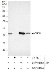
- Immunoprecipitation of TUFM protein from HepG2 whole cell extracts using 5 £gg of TUFM antibody (GTX101763) or TUFM antibody (GTX101764).Western blot analysis was performed using TUFM antibody (GTX101763).EasyBlot anti-Rabbit IgG (GTX221666-01) was used as a secondary reagent.
Supportive validation
- Submitted by
- GeneTex (provider)
- Main image

- Experimental details
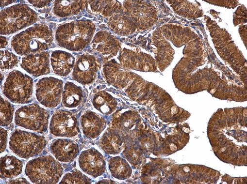
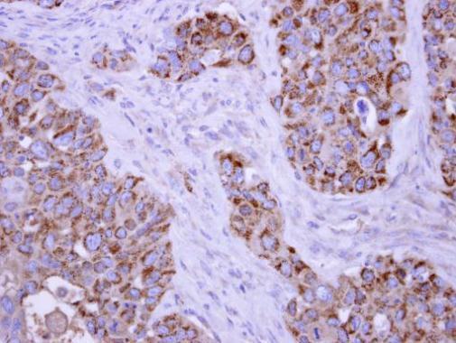
- TUFM antibody detects TUFM protein at mitochondria on H441 xenograft by immunohistochemical analysis. Sample: Paraffin-embedded H441 xenograft. TUFM antibody (GTX101763) dilution: 1:250.
- Submitted by
- GeneTex (provider)
- Main image
- Experimental details
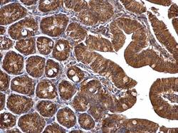
- TUFM antibody detects TUFM protein at mitochondria on mouse duodenum by immunohistochemical analysis. Sample: Paraffin-embedded mouse duodenum. TUFM antibody (GTX101763) dilution: 1:500.