 Explore
 Validate
 Learn
Learn Western blot
 Immunocytochemistry
Antibody data
- Antibody Data
- Antigen structure
- References [2]
- Comments [0]
- Validations
- Immunocytochemistry [1]
Submit
Validation data
Reference
Comment
Report error
- Product number
- HPA018991 - Provider product page

- Provider
- Atlas Antibodies
- Proper citation
- Atlas Antibodies Cat#HPA018991, RRID:AB_1858458
- Product name
- Anti-TUFM
- Antibody type
- Polyclonal
- Description
- Polyclonal Antibody against Human TUFM, Gene description: Tu translation elongation factor, mitochondrial, Alternative Gene Names: EF-TuMT, EFTu, Validated applications: WB, IHC, ICC, Uniprot ID: P49411, Storage: Store at +4°C for short term storage. Long time storage is recommended at -20°C.
- Reactivity
- Human
- Host
- Rabbit
- Conjugate
- Unconjugated
- Isotype
- IgG
- Vial size
- 100 µl
- Concentration
- 0.2 mg/ml
- Storage
- Store at +4°C for short term storage. Long time storage is recommended at -20°C.
- Handling
- The antibody solution should be gently mixed before use.
Submitted references Genetic disruption of slc4a10 alters the capacity for cellular metabolism and vectorial ion transport in the choroid plexus epithelium.
The Mitochondrial Proteins NLRX1 and TUFM Form a Complex that Regulates Type I Interferon and Autophagy
Christensen IB, Wu Q, Bohlbro AS, Skals MG, Damkier HH, Hübner CA, Fenton RA, Praetorius J
Fluids and barriers of the CNS 2020 Jan 7;17(1):2
Fluids and barriers of the CNS 2020 Jan 7;17(1):2
The Mitochondrial Proteins NLRX1 and TUFM Form a Complex that Regulates Type I Interferon and Autophagy
Lei Y, Wen H, Yu Y, Taxman D, Zhang L, Widman D, Swanson K, Wen K, Damania B, Moore C, Giguère P, Siderovski D, Hiscott J, Razani B, Semenkovich C, Chen X, Ting J
Immunity 2012;36(6):933-946
Immunity 2012;36(6):933-946
No comments: Submit comment
Supportive validation
- Submitted by
- Atlas Antibodies (provider)
- Main image
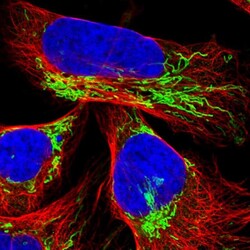
- Experimental details
- Immunofluorescent staining of human cell line U-2 OS shows localization to mitochondria.
- Sample type
- Human
