Explore
 Validate
 Learn
Learn Western blot
 Immunohistochemistry
Antibody data
- Antibody Data
- Antigen structure
- References [1]
- Comments [0]
- Validations
- Immunohistochemistry [1]
- Other assay [1]
Submit
Validation data
Reference
Comment
Report error
- Product number
- PA5-21849 - Provider product page

- Provider
- Invitrogen Antibodies
- Product name
- DRAK1 Polyclonal Antibody
- Antibody type
- Polyclonal
- Antigen
- Recombinant full-length protein
- Description
- Recommended positive controls: 293T, A431, H1299, HeLaS3, HepG2, Molt-4, Raji. Predicted reactivity: Rabbit (83%), Chicken (80%), Bovine (91%). Store product as a concentrated solution. Centrifuge briefly prior to opening the vial.
- Reactivity
- Human
- Host
- Rabbit
- Isotype
- IgG
- Vial size
- 100 μL
- Concentration
- 1.03 mg/mL
- Storage
- Store at 4°C short term. For long term storage, store at -20°C, avoiding freeze/thaw cycles.
Submitted references DAPK1 loss triggers tumor invasion in colorectal tumor cells.
Steinmann S, Kunze P, Hampel C, Eckstein M, Bertram Bramsen J, Muenzner JK, Carlé B, Ndreshkjana B, Kemenes S, Gasparini P, Friedrich O, Andersen C, Geppert C, Wang S, Eyupoglu I, Bäuerle T, Hartmann A, Schneider-Stock R
Cell death & disease 2019 Nov 26;10(12):895
Cell death & disease 2019 Nov 26;10(12):895
No comments: Submit comment
Supportive validation
- Submitted by
- Invitrogen Antibodies (provider)
- Main image
- Experimental details
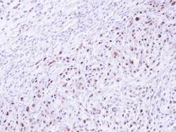
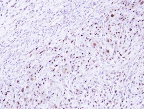
- Immunohistochemical analysis of paraffin-embedded SAS xenograft , using DRAK1 (Product # PA5-21849) antibody at 1:500 dilution. Antigen Retrieval: EDTA based buffer, pH 8.0, 15 min.
Supportive validation
- Submitted by
- Invitrogen Antibodies (provider)
- Main image
- Experimental details
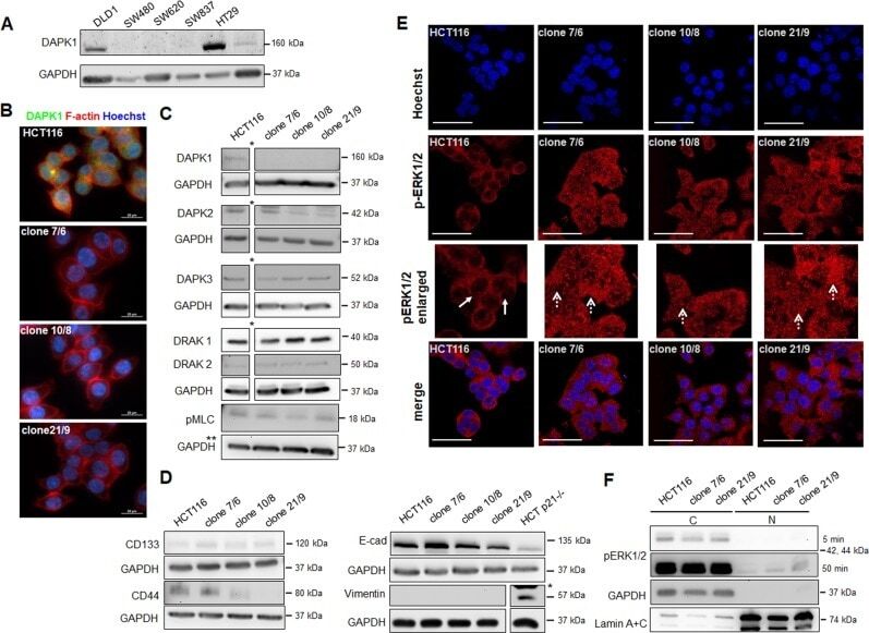
- Validation of CRISPR/Cas9-mediated DAPK1 knockout in HCT116 colorectal cancer cells. a Western Blot of DAPK1 expression status in various colorectal cancer cell lines. Representative images of two independent experiments are shown. GAPDH served as loading control. b Immunofluorescence staining of DAPK1 (green) in parental HCT116 cells and DAPK1 ko clones. Cells were counterstained with phalloidin for F-actin (red) and nuclear Hoechst (blue). Fluorescence microscopy was performed using a x 100 oil immersion objective. Representative images of two independent experiments are shown. Scale bar = 20 µm. c Protein expression of DAPK1 family members DAPK1, DAPK2, DAPK3, DRAK1, DRAK2 and DAPK1 phosphorylation target pMLC in HCT116 wildtype cells and DAPK1 ko clones were detected by Western Blotting using specific primary antibodies. Representative images of two independent experiments are shown. * images were cropped here; all samples were analyzed on the same SDS-PAGE gel; ** GAPDH blot has been used twice see Fig.5b, proteins have been loaded on the same membrane. d Western Blot analysis of stem cell markers (CD133, CD44) and EMT markers (epithelial marker: E-cad = E-cadherin, mesenchymal marker: Vimentin). Representative images of two independent experiments are shown.*images were cropped here; all samples were analyzed on the same SDS-PAGE gel. e Representative images of endogenous phospho-ERK1/2 (red) levels of immunostained HCT116 cells and DAPK1 ko clones examined by