Explore
 Validate
 Learn
Learn Western blot
 Immunocytochemistry
 Immunoprecipitation
Antibody data
- Antibody Data
- Antigen structure
- References [0]
- Comments [0]
- Validations
- Western blot [1]
- Immunocytochemistry [3]
Submit
Validation data
Reference
Comment
Report error
- Product number
- LS-C773643 - Provider product page

- Provider
- LSBio
- Product name
- BLVRA Antibody LS-C773643
- Antibody type
- Polyclonal
- Description
- Protein A purified
- Reactivity
- Human
- Host
- Rabbit
- Storage
- Store at -20°C.
No comments: Submit comment
Enhanced validation
- Submitted by
- LSBio (provider)
- Enhanced method
- Genetic validation
- Main image
- Experimental details
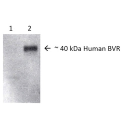
- Western blot analysis of Human, Rat Brain cell lysates showing detection of BVR protein using Rabbit Anti-BVR Polyclonal Antibody. Lane 1: Rat Brain. Lane 2: Human Brain lysates. Load: 10 µg. Primary Antibody: Rabbit Anti-BVR Polyclonal Antibody at 1:1000.

Supportive validation
- Submitted by
- LSBio (provider)
- Enhanced method
- Genetic validation
- Main image

- Experimental details
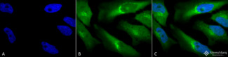
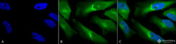
- Immunocytochemistry/Immunofluorescence analysis using Rabbit Anti-BVR Polyclonal Antibody. Tissue: Cervical cancer cell line (HeLa). Species: Human. Fixation: 2% Formaldehyde for 20 min at RT. Primary Antibody: Rabbit Anti-BVR Polyclonal Antibody at 1:120 for 12 hours at 4°C. Secondary Antibody: FITC Goat Anti-Rabbit (green) at 1:200 for 2 hours at RT. Counterstain: DAPI (blue) nuclear stain at 1:40000 for 2 hours at RT. Localization: Cytoplasm. Exosome. Magnification: 100x. (A) DAPI (blue) nuclear stain. (B) Anti-BVR Antibody. (C) Composite.

- Submitted by
- LSBio (provider)
- Main image
- Experimental details
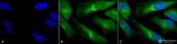
- Immunocytochemistry/Immunofluorescence analysis using Rabbit Anti-BVR Polyclonal Antibody. Tissue: Cervical cancer cell line (HeLa). Species: Human. Fixation: 2% Formaldehyde for 20 min at RT. Primary Antibody: Rabbit Anti-BVR Polyclonal Antibody at 1:120 for 12 hours at 4°C. Secondary Antibody: FITC Goat Anti-Rabbit (green) at 1:200 for 2 hours at RT. Counterstain: DAPI (blue) nuclear stain at 1:40000 for 2 hours at RT. Localization: Cytoplasm. Exosome. Magnification: 100x. (A) DAPI (blue) nuclear stain. (B) Anti-BVR Antibody. (C) Composite.
- Submitted by
- LSBio (provider)
- Main image
- Experimental details
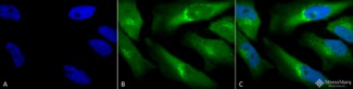
- Immunocytochemistry/Immunofluorescence analysis using Rabbit Anti-BVR Polyclonal Antibody. Tissue: Cervical cancer cell line (HeLa). Species: Human. Fixation: 2% Formaldehyde for 20 min at RT. Primary Antibody: Rabbit Anti-BVR Polyclonal Antibody at 1:120 for 12 hours at 4°C. Secondary Antibody: FITC Goat Anti-Rabbit (green) at 1:200 for 2 hours at RT. Counterstain: DAPI (blue) nuclear stain at 1:40000 for 2 hours at RT. Localization: Cytoplasm. Exosome. Magnification: 100x. (A) DAPI (blue) nuclear stain. (B) Anti-BVR Antibody. (C) Composite.