Explore
 Validate
 Learn
Learn Western blot
 Immunoprecipitation
Antibody data
- Antibody Data
- Antigen structure
- References [1]
- Comments [0]
- Validations
- Immunoprecipitation [1]
- Other assay [2]
Submit
Validation data
Reference
Comment
Report error
- Product number
- PA5-27340 - Provider product page

- Provider
- Invitrogen Antibodies
- Product name
- NFkB p100 Polyclonal Antibody
- Antibody type
- Polyclonal
- Antigen
- Recombinant full-length protein
- Description
- Recommended positive controls: A431, HeLa, HepG2, NIH-3T3 , YY-8103. Predicted reactivity: Mouse (91%), Rat (90%), Bovine (85%). Store product as a concentrated solution. Centrifuge briefly prior to opening the vial.
- Reactivity
- Human, Mouse
- Host
- Rabbit
- Isotype
- IgG
- Vial size
- 100 μL
- Concentration
- 1 mg/mL
- Storage
- Store at 4°C short term. For long term storage, store at -20°C, avoiding freeze/thaw cycles.
Submitted references IKKα regulates human keratinocyte migration through surveillance of the redox environment.
Lisse TS, Rieger S
Journal of cell science 2017 Mar 1;130(5):975-988
Journal of cell science 2017 Mar 1;130(5):975-988
No comments: Submit comment
Supportive validation
- Submitted by
- Invitrogen Antibodies (provider)
- Main image
- Experimental details
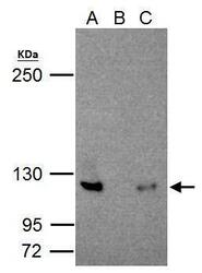
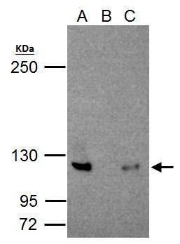
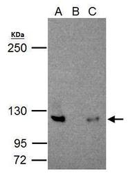
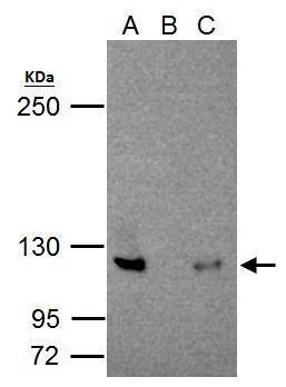
- NFkB p100/p52 antibody immunoprecipitates NFkB p100/p52 protein in IP experiments. IP Sample: Jurkat whole cell lysate/extract A. 30 µg Jurkat whole cell lysate/extract B. Control with 2 µg of preimmune rabbit IgG C. Immunoprecipitation of NFkB p100/p52 protein by 2 µg of NFkB p100/p52 antibody (Product # PA5-27340) 5% SDS-PAGE The immunoprecipitated NFkB p100/p52 protein was detected by NFkB p100/p52 antibody (Product # PA5-27340) diluted at 1:1,000.
Supportive validation
- Submitted by
- Invitrogen Antibodies (provider)
- Main image
- Experimental details
- NFkB p100/p52 antibody immunoprecipitates NFkB p100/p52 protein in IP experiments. IP Sample: Jurkat whole cell lysate/extract A. 30 µg Jurkat whole cell lysate/extract B. Control with 2 µg of preimmune rabbit IgG C. Immunoprecipitation of NFkB p100/p52 protein by 2 µg of NFkB p100/p52 antibody (Product # PA5-27340) 5% SDS-PAGE The immunoprecipitated NFkB p100/p52 protein was detected by NFkB p100/p52 antibody (Product # PA5-27340) diluted at 1:1,000.
- Submitted by
- Invitrogen Antibodies (provider)
- Main image
- Experimental details
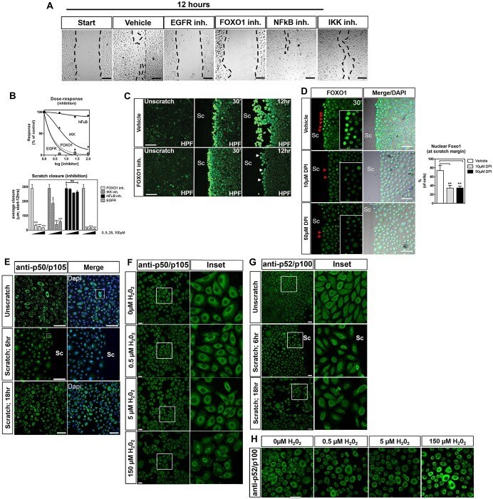
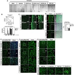
- Fig. 2. Scratch wound assay defines candidates for oxidation-dependent repair. (A) Effects of inhibitors on HEK001 scratch closure (5 uM sets shown). Inh., inhibitor. (B) Dose response of inhibitors measured using the scratch closure distance between the two edges of the wound. (C) HPF signals at the scratch margin in the absence and presence of FOXO1 inhibitor. SC, scratch. Time after scratching is shown in the top right. 30', 30 minutes. (D) Rapid nuclear localization of FOXO1 at the scratch margin of vehicle controls. Pre-treatment with DPI decreases FOXO1 staining at the scratch. Inset shows magnification of cells marked with red arrowheads. (E) Nuclear NF-kappaB1 (p50 and p105; p50/p105) is noticeable at the wound margin at 6 h post scratching but is cytoplasmic following wound closure at ~18 h, similar to in unscratched (unscratch) cells. (F) Predominantly cytoplasmic NF-kappaB1 p50/p105 staining was found in untreated cells and following H 2 O 2 treatment for 2 h. (G) Non-canonical NF-kappaB2 (p52 and p100; p52/p100) immunofluorescence following scratching shows predominantly cytoplasmic staining. (H) p52/p100 immunofluorescence reveals sustained cytoplasmic localization after H 2 O 2 treatment for 2 h. High H 2 O 2 concentrations lead to increased expression of NF-kappaB2. Scale bars: 100 um (A-E,H); 50 um (F,G). Two-way ANOVA and Bonferroni's multiple comparison post hoc tests were used. Significance: * P