Explore
 Validate
 Learn
Learn Western blot
Antibody data
- Antibody Data
- Antigen structure
- References [1]
- Comments [0]
- Validations
- Western blot [1]
- Immunocytochemistry [4]
- Immunohistochemistry [2]
Submit
Validation data
Reference
Comment
Report error
- Product number
- 701914 - Provider product page

- Provider
- Invitrogen Antibodies
- Product name
- alpha Actinin 2 Recombinant Rabbit Monoclonal Antibody (7H1L69)
- Antibody type
- Monoclonal
- Antigen
- Synthetic peptide
- Description
- This antibody is predicted to react with Monkey, Pig, Rat, Mouse Recombinant rabbit monoclonal antibodies are produced using in vitro expression systems. The expression systems are developed by cloning in the specific antibody DNA sequences from immunoreactive rabbits. Then, individual clones are screened to select the best candidates for production. The advantages of using recombinant rabbit monoclonal antibodies include: better specificity and sensitivity, lot-to-lot consistency, animal origin-free formulations, and broader immunoreactivity to diverse targets due to larger rabbit immune repertoire.
- Reactivity
- Human, Mouse, Rat
- Host
- Rabbit
- Isotype
- IgG
- Antibody clone number
- 7H1L69
- Vial size
- 100 μg
- Concentration
- 0.5 mg/mL
- Storage
- Store at 4°C short term. For long term storage, store at -20°C, avoiding freeze/thaw cycles.
Submitted references Deep learning detects cardiotoxicity in a high-content screen with induced pluripotent stem cell-derived cardiomyocytes.
Grafton F, Ho J, Ranjbarvaziri S, Farshidfar F, Budan A, Steltzer S, Maddah M, Loewke KE, Green K, Patel S, Hoey T, Mandegar MA
eLife 2021 Aug 2;10
eLife 2021 Aug 2;10
No comments: Submit comment
Supportive validation
- Submitted by
- Invitrogen Antibodies (provider)
- Main image
- Experimental details
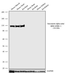
- Western blot analysis was performed on tissue extracts (30 µg lysate) of Mouse skeletal muscle (Lane 1), Mouse Heart (Lane 2), Rat Heart (Lane 3), Mouse Brain (Lane 4), Rat Brain (Lane 5), Mouse Kidney (Lane 6) and Rat Kidney (Lane 7). The blots were probed with Anti-Sarcomeric alpha actin/alpha actinin 2 Recombinant Rabbit Monoclonal Antibody (Product # 701914, 0.5 µg/mL). A 104 kDa band corresponding to Sarcomeric alpha actin/alpha actinin 2 was observed only in Muscle tissues. The blots were detected by chemiluminescence using Goat anti-Rabbit IgG (Heavy Chain) Superclonal™ Secondary Antibody, HRP conjugate (Product # A27036, 0.4 µg/mL, 1:5,000 dilution). Known quantity of protein samples were electrophoresed using Novex® NuPAGE® 4-12% Bis-Tris gel (Product # NP0321BOX), XCell SureLock™ Electrophoresis System (Product # EI0002) and Novex® Sharp Pre-Stained Protein Standard (Product # LC5800). Resolved proteins were then transferred onto a nitrocellulose membrane with iBlot® Dry Blotting System (Product # IB21001). The membrane was probed with the relevant primary and secondary Antibody following blocking with 5% skimmed milk. Chemiluminescent detection was performed using Pierce™ ECL Western blotting Substrate (Product # 32106).
Supportive validation
- Submitted by
- Invitrogen Antibodies (provider)
- Main image
- Experimental details
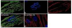
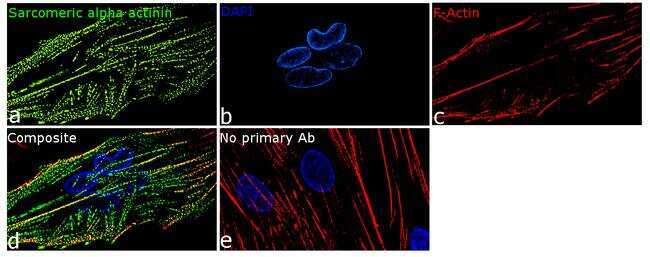
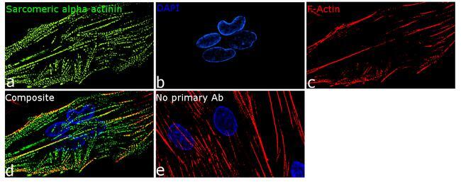
- For immunofluorescence analysis, HSkM cells were fixed and permeabilized for detection of endogenous sarcomeric alpha actinin using Anti-sarcomeric alpha actinin Recombinant Rabbit Monoclonal Antibody (Product # 701914, 2 µg/mL) and labeled with Goat anti-Rabbit IgG (H+L) Superclonal™ Secondary Antibody, Alexa Fluor® 488 conjugate (Product # A27034, 1:2,000). Panel a) shows representative cells that were stained for detection and localization of sarcomeric alpha actinin protein (green), Panel b) is stained for nuclei (blue) using SlowFade® Gold Antifade Mountant with DAPI (Product # S36938). Panel c) represents cytoskeletal F-actin staining using Rhodamine Phalloidin (Product # R415, 1:300). Panel d) is a composite image of Panels a, b and c clearly demonstrating cytoskeletal localization of sarcomeric alpha actinin. Panel e) represents control cells with no primary antibody to assess background. The images were captured at 60X magnification.
- Submitted by
- Invitrogen Antibodies (provider)
- Main image

- Experimental details
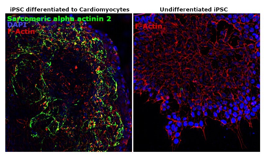
- For immunofluorescence analysis, iPSC differentiated to Cardiomyocytes were fixed and permeabilized for detection of endogenous alpha Actinin 2 using anti-Alpha actinin 2 Recombinant Rabbit monoclonal Antibody (Product # 701914, 1:100 dilution) and labeled with Goat anti-Rabbit IgG (H+L) Superclonal™ Secondary Antibody, Alexa Fluor® 488 conjugate (Product # A27034, 1:2,000). Nuclei (blue) is stained using ProLong™ Diamond Antifade Mountant with DAPI (Product # P36962). Panel c)and cytoskeletal F-actin stained using Rhodamine Phalloidin (Product # R415, 1:300). Panel a) shows undifferentiated iPSC cells, Panel b) is the image of differentiated cardiomyocytes showing cytoskeletal localization of sarcomeric alpha Actinin 2.

- Submitted by
- Invitrogen Antibodies (provider)
- Main image

- Experimental details
- For immunofluorescence analysis, HSkM cells were fixed and permeabilized for detection of endogenous sarcomeric alpha actinin using Anti-sarcomeric alpha actinin Recombinant Rabbit Monoclonal Antibody (Product # 701914, 2 µg/mL) and labeled with Goat anti-Rabbit IgG (Heavy Chain) Superclonal™ Secondary Antibody, Alexa Fluor® 488 conjugate (Product # A27034, 1:2,000). Panel a) shows representative cells that were stained for detection and localization of sarcomeric alpha actinin protein (green), Panel b) is stained for nuclei (blue) using SlowFade® Gold Antifade Mountant with DAPI (Product # S36938). Panel c) represents cytoskeletal F-actin staining using Rhodamine Phalloidin (Product # R415, 1:300). Panel d) is a composite image of Panels a, b and c clearly demonstrating cytoskeletal localization of sarcomeric alpha actinin. Panel e) represents control cells with no primary antibody to assess background. The images were captured at 60X magnification.
- Submitted by
- Invitrogen Antibodies (provider)
- Main image

- Experimental details
- For immunofluorescence analysis, iPSC differentiated to Cardiomyocytes were fixed and permeabilized for detection of endogenous alpha Actinin 2 using anti-Alpha actinin 2 Recombinant Rabbit monoclonal Antibody (Product # 701914, 1:100 dilution) and labeled with Goat anti-Rabbit IgG (Heavy Chain) Superclonal™ Secondary Antibody, Alexa Fluor® 488 conjugate (Product # A27034, 1:2,000). Nuclei (blue) is stained using ProLong™ Diamond Antifade Mountant with DAPI (Product # P36962). Panel c)and cytoskeletal F-actin stained using Rhodamine Phalloidin (Product # R415, 1:300). Panel a) shows undifferentiated iPSC cells, Panel b) is the image of differentiated cardiomyocytes showing cytoskeletal localization of sarcomeric alpha Actinin 2.
Supportive validation
- Submitted by
- Invitrogen Antibodies (provider)
- Main image
- Experimental details
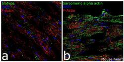
- Immunofluorescence analysis of Sarcomeric alpha actin in mouse heart tissue: Frozen sections were fixed with Acetone and blocked with 2% BSA. Whole heart longitudinal sections were then incubated with Anti- Sarcomeric alpha actin Recombinant Rabbit Monoclonal Antibody (Product # 701914, 2 µg/mL) overnight at 4°C, followed by Goat anti-Rabbit IgG (Heavy Chain) Superclonal™ Secondary Antibody, Alexa Fluor® 488 conjugate (Product # A27034, 1:2,000, 45 mins). Nuclei (blue) were stained using SlowFade® Gold Antifade Mountant with DAPI (Product # S36938), and cytoskeletal F-actin (red) was stained using Rhodamine Phalloidin (Product # R415, 1:300). Panel a) represents staining with the matched isotype control. Panel b) shows representative sections stained for Sarcomeric alpha actin (green). The images were captured at 20X magnification.

- Submitted by
- Invitrogen Antibodies (provider)
- Main image
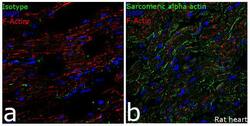
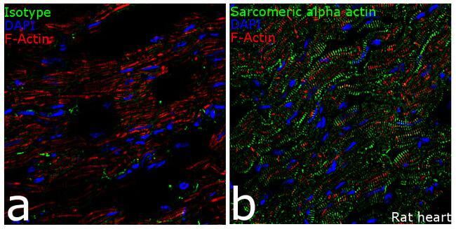
- Experimental details
- Immunofluorescence analysis of Sarcomeric alpha actin in Rat heart tissue: Frozen sections were fixed with Acetone and blocked with 2% BSA. Whole heart longitudinal sections were then incubated with Anti- Sarcomeric alpha actin Recombinant Rabbit Monoclonal Antibody (Product # 701914, 2 µg/mL) overnight at 4°C, followed by Goat anti-Rabbit IgG (Heavy Chain) Superclonal™ Secondary Antibody, Alexa Fluor® 488 conjugate (Product # A27034, 1:2,000, 45mins). Nuclei (blue) were stained using SlowFade® Gold Antifade Mountant with DAPI (Product # S36938), and cytoskeletal F-actin (red) was stained using Rhodamine Phalloidin (Product # R415, 1:300). Panel a) represents staining with the matched isotype control. Panel b) shows representative sections stained for Sarcomeric alpha actin (green). The images were captured at 20X magnification.