 Explore
 Validate
 Learn
Learn Western blot
Antibody data
- Antibody Data
- Antigen structure
- References [1]
- Comments [0]
- Validations
- Western blot [6]
- Immunocytochemistry [4]
- Immunohistochemistry [10]
Submit
Validation data
Reference
Comment
Report error
- Product number
- PA5-27863 - Provider product page

- Provider
- Invitrogen Antibodies
- Product name
- alpha Actinin 2 Polyclonal Antibody
- Antibody type
- Polyclonal
- Antigen
- Recombinant full-length protein
- Description
- Recommended positive controls: 293T, A431, HeLa, HepG2, Neuro2A, GL261, C8D30, NIH-3T3, Rat heart. Predicted reactivity: Mouse (100%), Rat (100%), Xenopus laevis (94%), Pig (99%), Chicken (98%), Bovine (99%). Store product as a concentrated solution. Centrifuge briefly prior to opening the vial.
- Reactivity
- Human, Mouse, Rat, Porcine
- Host
- Rabbit
- Isotype
- IgG
- Vial size
- 100 μL
- Concentration
- 0.18 mg/mL
- Storage
- Store at 4°C short term. For long term storage, store at -20°C, avoiding freeze/thaw cycles.
Submitted references Alterations in the host transcriptome in vitro following Rift Valley fever virus infection.
Pinkham C, Dahal B, de la Fuente CL, Bracci N, Beitzel B, Lindquist M, Garrison A, Schmaljohn C, Palacios G, Narayanan A, Campbell CE, Kehn-Hall K
Scientific reports 2017 Oct 30;7(1):14385
Scientific reports 2017 Oct 30;7(1):14385
No comments: Submit comment
Supportive validation
- Submitted by
- Invitrogen Antibodies (provider)
- Main image
- Experimental details
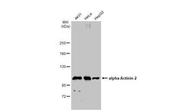
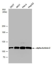
- Western Blot using alpha Actinin 2 Polyclonal Antibody (Product # PA5-27863). Various whole cell extracts (30 µg) were separated by 5% SDS-PAGE, and the membrane was blotted with alpha Actinin 2 Polyclonal Antibody (Product # PA5-27863) diluted at 1:1,000. The HRP-conjugated anti-rabbit IgG antibody was used to detect the primary antibody.
- Submitted by
- Invitrogen Antibodies (provider)
- Main image
- Experimental details
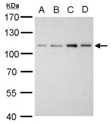
- alpha Actinin 2 Polyclonal Antibody detects ACTN2 protein by western blot analysis. A. 30 µg Neuro2A whole cell lysate/extract. B. 30 µg GL261 whole cell lysate/extract. C. 30 µg C8D30 whole cell lysate/extract. D. 30 µg NIH-3T3 whole cell lysate/extract.7.5% SDS-PAGE. Alpha Actinin 2 Polyclonal Antibody (Product # PA5-27863) dilution: 1:1,000. The HRP-conjugated anti-rabbit IgG antibody was used to detect the primary antibody.

- Submitted by
- Invitrogen Antibodies (provider)
- Main image
- Experimental details
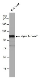
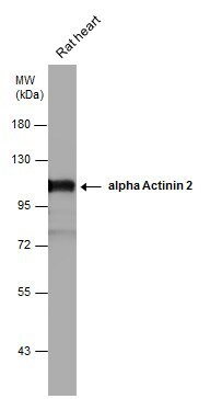
- alpha Actinin 2 antibody detects alpha Actinin 2 protein by western blot analysis. Rat tissue extracts (50 µg) was separated by 7.5% SDS-PAGE, and the membrane was blotted with alpha Actinin 2 antibody alpha Actinin 2 Polyclonal Antibody (Product # PA5-27863) diluted at 1:5,000. The HRP-conjugated anti-rabbit IgG antibody was used to detect the primary antibody.
- Submitted by
- Invitrogen Antibodies (provider)
- Main image
- Experimental details
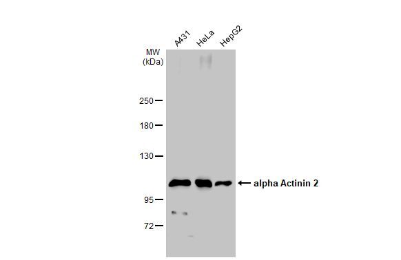
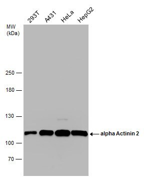
- Western Blot analysis of alpha Actinin 2 was performed by separating 30 µg of various whole cell extracts by 5% SDS-PAGE. Proteins were transferred to a membrane and probed with a alpha Actinin 2 Polyclonal Antibody (Product # PA5-27863) at a dilution of 1:1000.
- Submitted by
- Invitrogen Antibodies (provider)
- Main image
- Experimental details
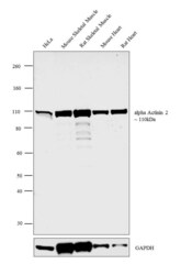
- Western blot analysis was performed on whole cell extract (30 µg lysate) of HeLa (Lane 1), tissue extracts (30 µg lysate) of Mouse Skeletal Muscle (Lane 2), Rat Skeletal Muscle (Lane 3), Mouse Heart (Lane 4) and Rat Heart (Lane 5). The blot was probed with Anti-alpha Actinin 2 Polyclonal Antibody (Product # PA5-27863, 1:500 dilution) and detected by chemiluminescence using Goat anti-Rabbit IgG (Heavy Chain) Superclonal™ Secondary Antibody, HRP conjugate (Product # A27036, 0.25 µg/mL, 1:4000 dilution). A 110 kDa band corresponding to alpha Actinin 2 was detected across the cell line and tissues tested.

- Submitted by
- Invitrogen Antibodies (provider)
- Main image

- Experimental details
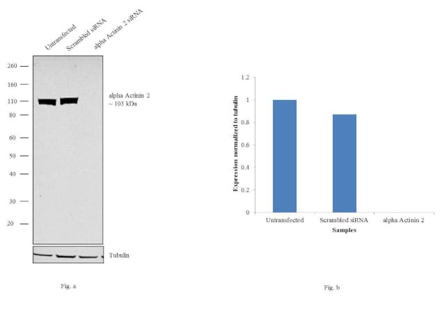
- Knockdown of Alpha Actinin 2 was achieved by transfecting HeLa cells with Alpha Actinin 2 specific siRNAs (Silencer® select Product # s32, s30). Western blot analysis (Fig. a) was performed using whole cell extracts from the Alpha Actinin 2 knockdown cells (lane 3), non-specific scrambled siRNA transfected cells (lane 2) and untransfected cells (lane 1). The blots were probed with Alpha Actinin 2 Polyclonal Antibody (Product # PA5-27863, 1:1500 dilution) and Goat anti-Rabbit IgG (Heavy Chain) Superclonal™ Secondary Antibody, HRP conjugate (Product # A27036, 0.25 µg/mL, 1:4000 dilution). Densitometric analysis of this western blot is shown in histogram (Fig. b). Decrease in signal upon siRNA mediated knock down confirms that antibody is specific to Alpha Actinin 2.
Supportive validation
- Submitted by
- Invitrogen Antibodies (provider)
- Main image

- Experimental details
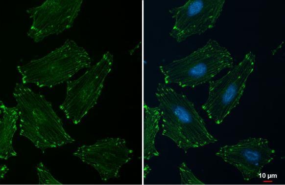
- Immunocytochemistry-Immunofluorescence analysis of alpha Actinin 2 was performed in HeLa cells fixed in ice cold MeOH for 5 min. Green: alpha Actinin 2 Polyclonal Antibody (Product # PA5 27863) diluted at 1:500. Blue: Hoechst 33342 staining.

- Submitted by
- Invitrogen Antibodies (provider)
- Main image

- Experimental details
- alpha Actinin 2 Polyclonal Antibody detects alpha Actinin 2 protein at cytoskeleton by immunofluorescent analysis. Sample: HeLa cells were fixed in ice-cold MeOH for 5 min. Green: alpha Actinin 2 stained by alpha Actinin 2 Polyclonal Antibody (Product # PA5-27863) diluted at 1:500. Blue: Fluoroshield with DAPI .

- Submitted by
- Invitrogen Antibodies (provider)
- Main image

- Experimental details
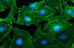
- alpha Actinin 2 Polyclonal Antibody detects alpha Actinin 2 protein at cytoskeleton by immunofluorescent analysis. Sample: HeLa cells were fixed in ice-cold MeOH for 5 min. Green: alpha Actinin 2 stained by alpha Actinin 2 Polyclonal Antibody (Product # PA5-27863) diluted at 1:500. Blue: Fluoroshield with DAPI .
- Submitted by
- Invitrogen Antibodies (provider)
- Main image
- Experimental details
- Immunocytochemistry-Immunofluorescence analysis of alpha Actinin 2 was performed in HeLa cells fixed in ice cold MeOH for 5 min. Green: alpha Actinin 2 Polyclonal Antibody (Product # PA5 27863) diluted at 1:500. Blue: Hoechst 33342 staining.

Supportive validation
- Submitted by
- Invitrogen Antibodies (provider)
- Main image

- Experimental details
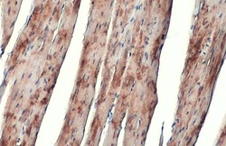
- Immunohistochemistry (Paraffin) analysis of alpha Actinin 2 was performed in paraffin-embedded mouse heart tissue using alpha Actinin 2 Polyclonal Antibody (Product # PA5-27863) at a dilution of 1:2000. Antigen Retrieval: Citrate buffer, pH 6.0, 15 min.

- Submitted by
- Invitrogen Antibodies (provider)
- Main image

- Experimental details
- alpha Actinin 2 Polyclonal Antibody detects alpha Actinin 2 protein at cytoplasm by immunohistochemical analysis. Sample: Paraffin-embedded mouse heart. alpha Actinin 2 stained by alpha Actinin 2 Polyclonal Antibody (Product # PA5-27863) diluted at 1:500. Antigen Retrieval: Citrate buffer, pH 6.0, 15 min.

- Submitted by
- Invitrogen Antibodies (provider)
- Main image

- Experimental details
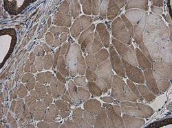
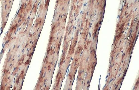
- alpha Actinin 2 Polyclonal Antibody detects alpha Actinin 2 protein at cytoplasm by immunohistochemical analysis. Sample: Paraffin-embedded mouse muscle. alpha Actinin 2 stained by alpha Actinin 2 Polyclonal Antibody (Product # PA5-27863) diluted at 1:500. Antigen Retrieval: Citrate buffer, pH 6.0, 15 min.

- Submitted by
- Invitrogen Antibodies (provider)
- Main image
- Experimental details
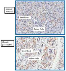
- Immunohistochemistry analysis of Actinin 2 was performed on normal (top) and chronic pancreatitis (bottom) human tissue. To expose target proteins, antigen retrieval was performed by microwaving tissues for 8-15 minutes in 10mM sodium citrate buffer (pH 6.0). Following antigen retrieval, endogenous peroxidases were blocked with 3% hydrogen peroxide-methanol for 15 min at room temperature. Tissue slides were washed with deionized water and PBS, and then blocked in 3% BSA-PBS for 30 min at room temperature. Tissues were probed with a ACTN2 polyclonal antibody (Product # PA5-27863) diluted 1:500 in 3% BSA-PBS overnight at 4°C in a humidified chamber. Tissues were washed extensively in PBST and detection was performed using an HRP-conjugated secondary antibody followed by colorimetric detection using a DAB kit. Tissues were counterstained with hematoxylin and dehydrated with ethanol and xylene to prep for mounting. Images were taken at 20X magnification. Data courtesy of Antibody Data Exchange Program.

- Submitted by
- Invitrogen Antibodies (provider)
- Main image
- Experimental details
- Immunohistochemistry (Paraffin) analysis of alpha Actinin 2 was performed in paraffin-embedded mouse muscle tissue using alpha Actinin 2 Polyclonal Antibody (Product # PA5-27863) at a dilution of 1:500.

- Submitted by
- Invitrogen Antibodies (provider)
- Main image

- Experimental details
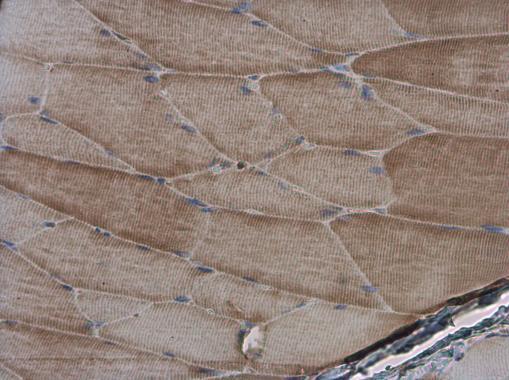
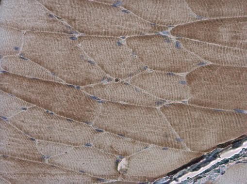
- Immunohistochemistry (Paraffin) analysis of alpha Actinin 2 was performed in paraffin-embedded rat muscle tissue using alpha Actinin 2 Polyclonal Antibody (Product # PA5-27863) at a dilution of 1:500.
- Submitted by
- Invitrogen Antibodies (provider)
- Main image
- Experimental details
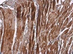
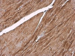
- Immunohistochemistry (Paraffin) analysis of alpha Actinin 2 was performed in paraffin-embedded rat heart tissue using alpha Actinin 2 Polyclonal Antibody (Product # PA5-27863) at a dilution of 1:2000. Antigen Retrieval: Citrate buffer, pH 6.0, 15 min.

- Submitted by
- Invitrogen Antibodies (provider)
- Main image
- Experimental details
- alpha Actinin 2 Polyclonal Antibody detects alpha Actinin 2 protein at cytoplasm by immunohistochemical analysis. Sample: Paraffin-embedded mouse heart. alpha Actinin 2 stained by alpha Actinin 2 Polyclonal Antibody (Product # PA5-27863) diluted at 1:500. Antigen Retrieval: Citrate buffer, pH 6.0, 15 min.
- Submitted by
- Invitrogen Antibodies (provider)
- Main image

- Experimental details
- Immunohistochemistry (Paraffin) analysis of alpha Actinin 2 was performed in paraffin-embedded rat muscle tissue using alpha Actinin 2 Polyclonal Antibody (Product # PA5-27863) at a dilution of 1:500.
- Submitted by
- Invitrogen Antibodies (provider)
- Main image
- Experimental details
- Immunohistochemistry (Paraffin) analysis of alpha Actinin 2 was performed in paraffin-embedded mouse heart tissue using alpha Actinin 2 Polyclonal Antibody (Product # PA5-27863) at a dilution of 1:2000. Antigen Retrieval: Citrate buffer, pH 6.0, 15 min.
