Explore
 Validate
 Learn
Learn Western blot
Antibody data
- Antibody Data
- Antigen structure
- References [1]
- Comments [0]
- Validations
- Western blot [1]
- Immunohistochemistry [8]
Submit
Validation data
Reference
Comment
Report error
- Product number
- OSR00190W - Provider product page

- Provider
- Invitrogen Antibodies
- Product name
- TRPV2 Polyclonal Antibody
- Antibody type
- Polyclonal
- Antigen
- Synthetic peptide
- Description
- Reconstitute lyophilized product with 1 mL distilled water.
- Reactivity
- Human, Mouse, Rat
- Host
- Rabbit
- Isotype
- IgG
- Vial size
- 100 µL
- Concentration
- Conc. Not Determined
- Storage
- Store at 4°C short term. For long term storage, store at -20°C, avoiding freeze/thaw cycles. Glycerol (1:1) may be added for added stability.
Submitted references Evaluation of cationic channel TRPV2 as a novel biomarker and therapeutic target in Leukemia-Implications concerning the resolution of pulmonary inflammation.
Siveen KS, Prabhu KS, Parray AS, Merhi M, Arredouani A, Chikri M, Uddin S, Dermime S, Mohammad RM, Steinhoff M, Janahi IA, Azizi F
Scientific reports 2019 Feb 7;9(1):1554
Scientific reports 2019 Feb 7;9(1):1554
No comments: Submit comment
Supportive validation
- Submitted by
- Invitrogen Antibodies (provider)
- Main image

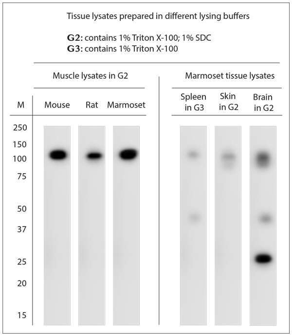
- Experimental details
- Western blot analysis was performed on tissue lysates using TRPV2 Polyclonal Antibody (Product #OSR00190W). Blocking: 1% LFDM for 30 min at RT; primary antibody dilutions: in Muscle 1:4000; in Brain, Spleen, Skin: 1:3000; incubated at 4C overnight.
Supportive validation
- Submitted by
- Invitrogen Antibodies (provider)
- Main image
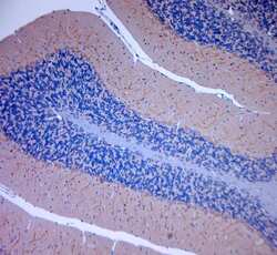
- Experimental details
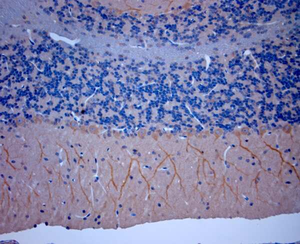
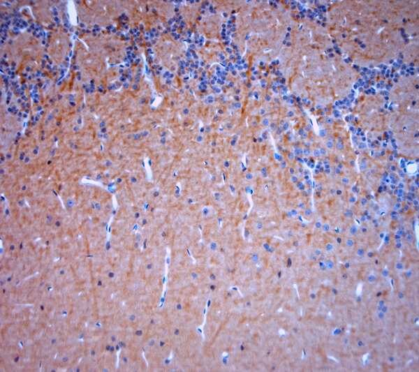
- Immunohistochemistry (Paraffin) analysis was performed in sections of mouse cerebellum using TRPV2 Polyclonal Antibody (Product #OSR00190W). The animal was perfused using Autoperfuser at a pressure of 130 mmHg with 300 ml 4% FA being processed for paraffin embedding. HIER: Tris-EDTA, pH 9 for 20 min using Thermo PT Module. Blocking: 0.2% LFDM in TBST filtered thru 0.2 µm. Primary antibody: dilution 1:2000, incubated 30 min at RT using Autostainer. Sections were counterstained with Harris Hematoxylin.

- Submitted by
- Invitrogen Antibodies (provider)
- Main image
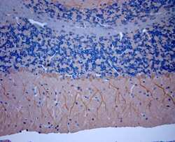
- Experimental details
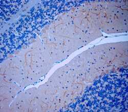
- Immunohistochemistry (Paraffin) analysis was performed in sections of mouse cerebellum using TRPV2 Polyclonal Antibody (Product #OSR00190W). The animal was perfused using Autoperfuser at a pressure of 130 mmHg with 300 ml 4% FA being processed for paraffin embedding. HIER: Tris-EDTA, pH 9 for 20 min using Thermo PT Module. Blocking: 0.2% LFDM in TBST filtered thru 0.2 µm. Primary antibody: dilution 1:2000, incubated 30 min at RT using Autostainer. Sections were counterstained with Harris Hematoxylin.
- Submitted by
- Invitrogen Antibodies (provider)
- Main image
- Experimental details
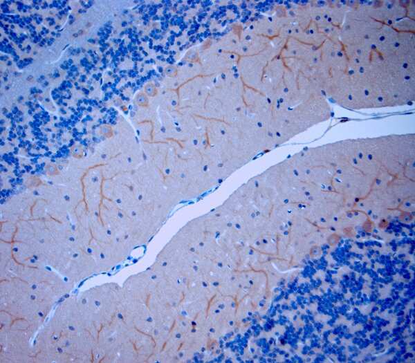
- Immunohistochemistry (Paraffin) analysis was performed in sections of mouse cerebellum using TRPV2 Polyclonal Antibody (Product #OSR00190W). The animal was perfused using Autoperfuser at a pressure of 130 mmHg with 300 ml 4% FA being processed for paraffin embedding. HIER: Tris-EDTA, pH 9 for 20 min using Thermo PT Module. Blocking: 0.2% LFDM in TBST filtered thru 0.2 µm. Primary antibody: dilution 1:2000, incubated 30 min at RT using Autostainer. Sections were counterstained with Harris Hematoxylin.
- Submitted by
- Invitrogen Antibodies (provider)
- Main image
- Experimental details
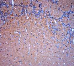
- Immunohistochemistry (Paraffin) analysis was performed in sections of mouse olfactory bulbs using TRPV2 Polyclonal Antibody (Product #OSR00190W). The animal was perfused using Autoperfuser at a pressure of 130 mmHg with 300 ml 4% FA being processed for paraffin embedding. HIER: Tris-EDTA, pH 9 for 20 min using Thermo PT Module. Blocking: 0.2% LFDM in TBST filtered thru 0.2 µm. Primary antibody: dilution 1:2000, incubated 30 min at RT using Autostainer. Sections were counterstained with Harris Hematoxylin.
- Submitted by
- Invitrogen Antibodies (provider)
- Main image
- Experimental details
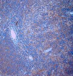
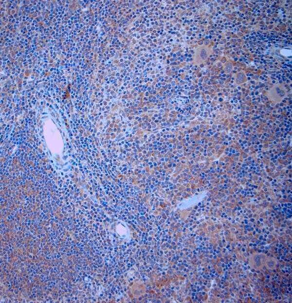
- Immunohistochemistry (Paraffin) analysis was performed in sections of mouse spleen using TRPV2 Polyclonal Antibody (Product #OSR00190W). The animal was perfused using Autoperfuser at a pressure of 130 mmHg with 300 ml 4% FA being processed for paraffin embedding. HIER: Tris-EDTA, pH 9 for 20 min using Thermo PT Module. Blocking: 0.2% LFDM in TBST filtered thru 0.2 µm. Primary antibody: dilution 1:2000, incubated 30 min at RT using Autostainer. Sections were counterstained with Harris Hematoxylin.
- Submitted by
- Invitrogen Antibodies (provider)
- Main image
- Experimental details
- Immunohistochemistry (Paraffin) analysis was performed in sections of mouse DRG using TRPV2 Polyclonal Antibody (Product #OSR00190W). The animal was perfused using Autoperfuser at a pressure of 130 mmHg with 300 ml 4% FA being processed for paraffin embedding. HIER: Tris-EDTA, pH 9 for 20 min using Thermo PT Module. Blocking: 0.2% LFDM in TBST filtered thru 0.2 µm. Primary antibody: dilution 1:2000, incubated 30 min at RT using Autostainer. Sections were counterstained with Harris Hematoxylin.

- Submitted by
- Invitrogen Antibodies (provider)
- Main image
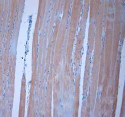
- Experimental details
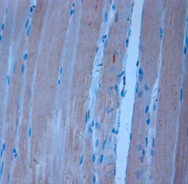
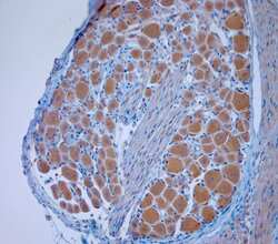
- Immunohistochemistry (Paraffin) analysis was performed in sections of rat muscle using TRPV2 Polyclonal Antibody (Product #OSR00190W). The animal was perfused using Autoperfuser at a pressure of 130 mmHg with 300 ml 4% FA being processed for paraffin embedding. HIER: Tris-EDTA, pH 9 for 20 min using Thermo PT Module. Blocking: 0.2% LFDM in TBST filtered thru 0.2 µm. Primary antibody: dilution 1:2000, incubated 30 min at RT using Autostainer. Sections were counterstained with Harris Hematoxylin.
- Submitted by
- Invitrogen Antibodies (provider)
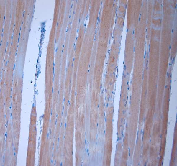
- Main image

- Experimental details
- Immunohistochemistry (Paraffin) analysis was performed in sections of rat muscle using TRPV2 Polyclonal Antibody (Product #OSR00190W). The animal was perfused using Autoperfuser at a pressure of 130 mmHg with 300 ml 4% FA being processed for paraffin embedding. HIER: Tris-EDTA, pH 9 for 20 min using Thermo PT Module. Blocking: 0.2% LFDM in TBST filtered thru 0.2 µm. Primary antibody: dilution 1:2000, incubated 30 min at RT using Autostainer. Sections were counterstained with Harris Hematoxylin.