Explore
 Validate
 Learn
LearnMA1-199
antibody from Invitrogen Antibodies
Targeting: HNF4A
HNF4, MODY, MODY1, NR2A1, TCF14
 Western blot
 ELISA
ELISA Immunocytochemistry Immunoprecipitation Immunohistochemistry Flow cytometry Other assay
Antibody data
- Antibody Data
- Antigen structure
- References [7]
- Comments [0]
- Validations
- Immunocytochemistry [6]
- Flow cytometry [2]
- Other assay [6]
Submit
Validation data
Reference
Comment
Report error
- Product number
- MA1-199 - Provider product page

- Provider
- Invitrogen Antibodies
- Product name
- HNF4A Monoclonal Antibody (K9218)
- Antibody type
- Monoclonal
- Antigen
- Recombinant full-length protein
- Description
- MA1-199 detects HNF4a in human, mouse and rat samples.
- Reactivity
- Human, Mouse, Rat
- Host
- Mouse
- Isotype
- IgG
- Antibody clone number
- K9218
- Vial size
- 100 μL
- Concentration
- 1 mg/mL
- Storage
- Maintain refrigerated at 2-8°C for up to 1 month. For long term storage store at -20°C
Submitted references SARS-CoV-2 infection of human pluripotent stem cell-derived liver organoids reveals potential mechanisms of liver pathology.
In vitro grafting of hepatic spheroids and organoids on a microfluidic vascular bed.
DevKidCC allows for robust classification and direct comparisons of kidney organoid datasets.
Establishment of Human Leukocyte Antigen-Mismatched Immune Responses after Transplantation of Human Liver Bud in Humanized Mouse Models.
Enhanced expression of HNF4α during intestinal epithelial differentiation is involved in the activation of ER stress.
An Unexpected Role of Cholesterol Sulfotransferase and its Regulation in Sensitizing Mice to Acetaminophen-Induced Liver Injury.
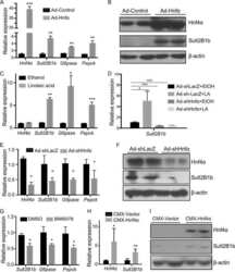
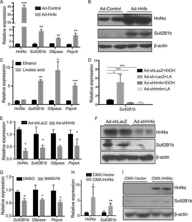
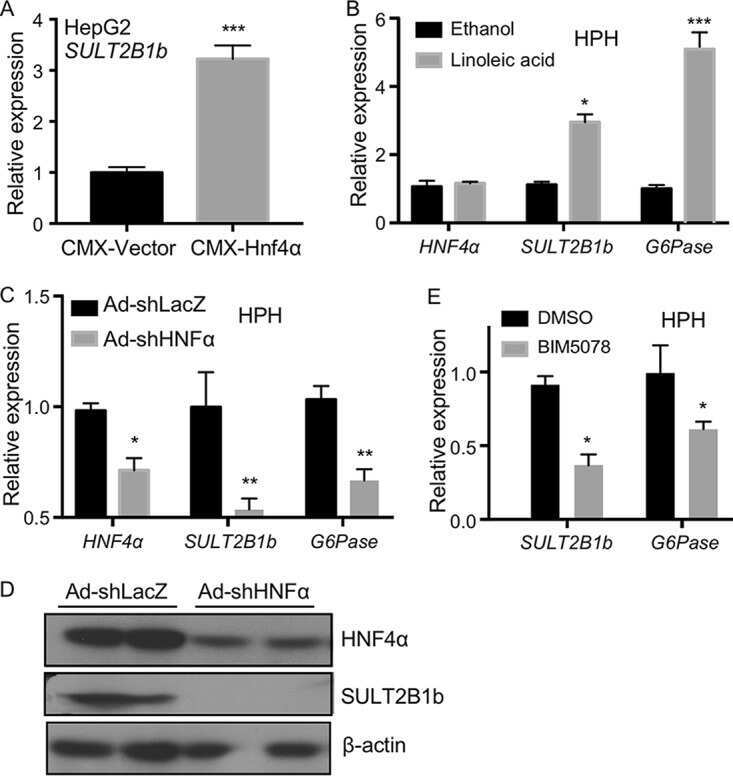
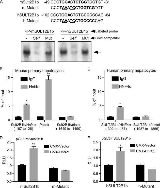
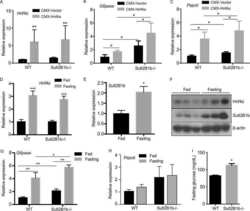
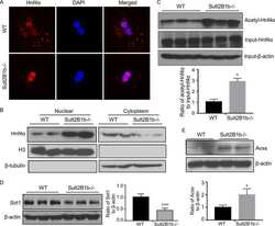
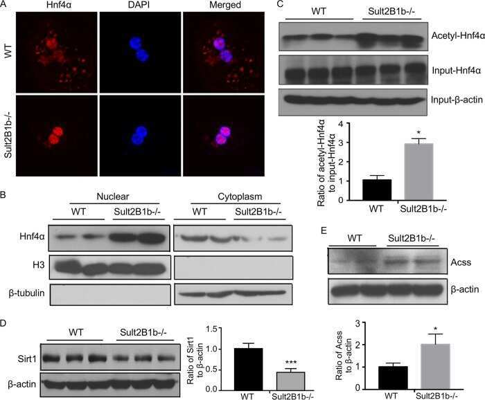
Regulation of Cholesterol Sulfotransferase SULT2B1b by Hepatocyte Nuclear Factor 4α Constitutes a Negative Feedback Control of Hepatic Gluconeogenesis.
Richards A, Friesen M, Khalil A, Barrasa MI, Gehrke L, Jaenisch R
iScience 2022 Oct 21;25(10):105146
iScience 2022 Oct 21;25(10):105146
In vitro grafting of hepatic spheroids and organoids on a microfluidic vascular bed.
Bonanini F, Kurek D, Previdi S, Nicolas A, Hendriks D, de Ruiter S, Meyer M, Clapés Cabrer M, Dinkelberg R, García SB, Kramer B, Olivier T, Hu H, López-Iglesias C, Schavemaker F, Walinga E, Dutta D, Queiroz K, Domansky K, Ronden B, Joore J, Lanz HL, Peters PJ, Trietsch SJ, Clevers H, Vulto P
Angiogenesis 2022 Nov;25(4):455-470
Angiogenesis 2022 Nov;25(4):455-470
DevKidCC allows for robust classification and direct comparisons of kidney organoid datasets.
Wilson SB, Howden SE, Vanslambrouck JM, Dorison A, Alquicira-Hernandez J, Powell JE, Little MH
Genome medicine 2022 Feb 22;14(1):19
Genome medicine 2022 Feb 22;14(1):19
Establishment of Human Leukocyte Antigen-Mismatched Immune Responses after Transplantation of Human Liver Bud in Humanized Mouse Models.
Mori A, Murata S, Tashiro N, Tadokoro T, Okamoto S, Otsuka R, Wada H, Murata T, Takahashi T, Seino KI, Taniguchi H
Cells 2021 Feb 23;10(2)
Cells 2021 Feb 23;10(2)
Enhanced expression of HNF4α during intestinal epithelial differentiation is involved in the activation of ER stress.
Tunçer S, Sade-Memişoğlu A, Keşküş AG, Sheraj I, Güner G, Akyol A, Banerjee S
The FEBS journal 2020 Jun;287(12):2504-2523
The FEBS journal 2020 Jun;287(12):2504-2523
An Unexpected Role of Cholesterol Sulfotransferase and its Regulation in Sensitizing Mice to Acetaminophen-Induced Liver Injury.
An Y, Wang P, Xu P, Tung HC, Xie Y, Kirisci L, Xu M, Ren S, Tian X, Ma X, Xie W
Molecular pharmacology 2019 Jun;95(6):597-605
Molecular pharmacology 2019 Jun;95(6):597-605
Regulation of Cholesterol Sulfotransferase SULT2B1b by Hepatocyte Nuclear Factor 4α Constitutes a Negative Feedback Control of Hepatic Gluconeogenesis.
Bi Y, Shi X, Zhu J, Guan X, Garbacz WG, Huang Y, Gao L, Yan J, Xu M, Ren S, Ren S, Liu Y, Ma X, Li S, Xie W
Molecular and cellular biology 2018 Apr 1;38(7)
Molecular and cellular biology 2018 Apr 1;38(7)
No comments: Submit comment
Supportive validation
- Submitted by
- Invitrogen Antibodies (provider)
- Main image
- Experimental details
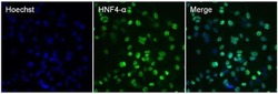
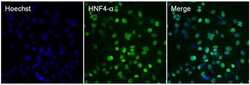
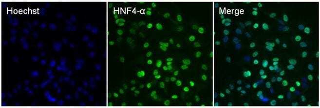
- Immunofluorescent analysis of HNF4 alpha (green) in HepG2 cells. The cells were fixed with 4% paraformaldehyde for 15 minutes, permeabilized with 0.1% Triton X-100 in PBS for 15 minutes, and blocked with 0.3% BSA in PBS (Product # 37525) for 30 minutes at room temperature. Cells were stained with a HNF4 alpha mouse monoclonal antibody (Product # MA1-199) at a dilution of 2.5 µg/mL in staining buffer for 1 hour at room temperature, and then incubated with a Goat anti-Mouse IgG Secondary Antibody, DyLight 488 conjugate (Product # 35503) at a dilution of 1:500 for 1 hour at room temperature (green). Nuclei (blue) were counterstained with Hoechst 33342 dye (Product # 62249). Images were taken on a Thermo Scientific ToxInsight Instrument at 20X magnification.
- Submitted by
- Invitrogen Antibodies (provider)
- Main image
- Experimental details
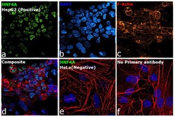
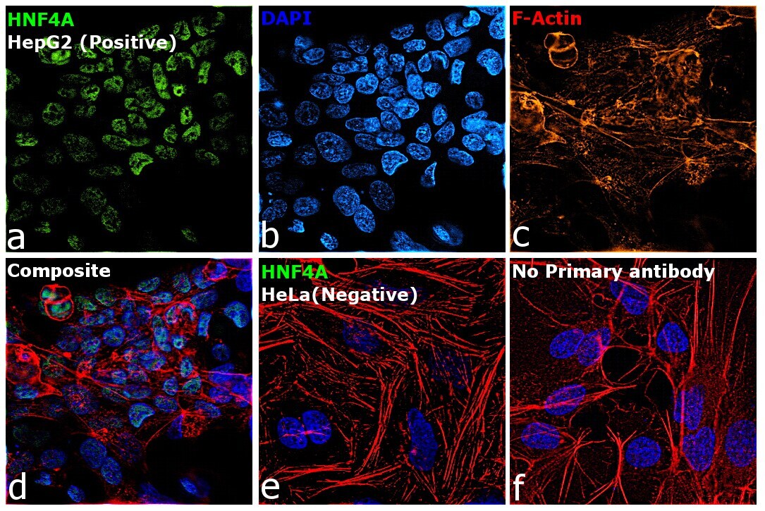
- Immunofluorescence analysis of HNF4A was performed using 70% confluent log phase HepG2 and HeLa cells. The cells were fixed with 4% Paraformaldehyde for 10 minutes, permeabilized with 0.1% Triton™ X-100 for 10 minutes, and blocked with 2% BSA for 10 minutes at room temperature. The cells were labeled with Anti-HNF4A Monoclonal Antibody (Product # MA1-199) at 1:100 dilution in 0.1% BSA, incubated at 4 degree celsius overnight and then labeled with Goat anti-Mouse IgG (H+L) Superclonal™ Recombinant Secondary Antibody, Alexa Fluor® 488 conjugate (Product # A28175, 1:2000 dilution) for 45 minutes at room temperature (Panel a: Green). Nuclei (Panel b: Blue) were stained with SlowFade® Gold Antifade Mountant with DAPI (Product # S36938). F-actin (Panel c: Red) was stained with Rhodamine Phalloidin (Product # R415, 1:300). Panel d represents the merged image showing nuclear localization. Panel e represents HeLa cells having no expression of HNF4A. Panel f represents control cells with no primary antibody to assess background. The images were captured at 60X magnification.

- Submitted by
- Invitrogen Antibodies (provider)
- Main image
- Experimental details
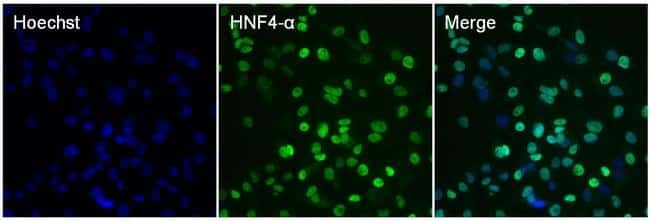
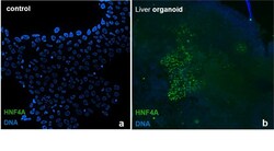
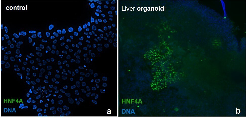
- Immunofluorescence analysis of HNF4A was performed in iPSC derived liver organoid. The cells were fixed with 4% Paraformaldehyde for 30 minutes, permeabilized with 0.1% Triton™ X-100 for 15 minutes and blocked with 2% BSA for 1 hour at room temperature. The cells were labeled with Anti-HNF4A Monoclonal Antibody (Product # MA1-199) at 1:100 dilution in 0.1% BSA, incubated at 4°C overnight and then labeled with Goat anti-Mouse IgG (H+L) Superclonal™ Recombinant Secondary Antibody, Alexa Fluor® 488 conjugate (Product # A28175, 1:2,000 dilution) for 45 minutes at room temperature. The panels represent the merged images of HNF4A Monoclonal Antibody (green) and DAPI (blue) staining for iPSC control cells and differentiated liver organoid. The images were captured at 20X magnification.

- Submitted by
- Invitrogen Antibodies (provider)
- Main image

- Experimental details
- Immunofluorescence analysis of HNF4A was performed in iPSC derived liver organoid. The cells were fixed with 4% Paraformaldehyde for 30 minutes, permeabilized with 0.1% Triton™ X-100 for 15 minutes and blocked with 2% BSA for 1 hour at room temperature. The cells were labeled with Anti-HNF4A Monoclonal Antibody (Product # MA1-199) at 1:100 dilution in 0.1% BSA, incubated at 4°C overnight and then labeled with Goat anti-Mouse IgG (H+L) Superclonal™ Recombinant Secondary Antibody, Alexa Fluor® 488 conjugate (Product # A28175, 1:2,000 dilution) for 45 minutes at room temperature. The panels represent the merged images of HNF4A Monoclonal Antibody (green) and DAPI (blue) staining for iPSC control cells and differentiated liver organoid. The images were captured at 20X magnification.
- Submitted by
- Invitrogen Antibodies (provider)
- Main image

- Experimental details
- Immunofluorescence analysis of HNF4A was performed using 70% confluent log phase HepG2 and HeLa cells. The cells were fixed with 4% Paraformaldehyde for 10 minutes, permeabilized with 0.1% Triton™ X-100 for 10 minutes, and blocked with 2% BSA for 10 minutes at room temperature. The cells were labeled with Anti-HNF4A Monoclonal Antibody (Product # MA1-199) at 1:100 dilution in 0.1% BSA, incubated at 4 degree celsius overnight and then labeled with Goat anti-Mouse IgG (H+L) Superclonal™ Recombinant Secondary Antibody, Alexa Fluor® 488 conjugate (Product # A28175, 1:2000 dilution) for 45 minutes at room temperature (Panel a: Green). Nuclei (Panel b: Blue) were stained with SlowFade® Gold Antifade Mountant with DAPI (Product # S36938). F-actin (Panel c: Red) was stained with Rhodamine Phalloidin (Product # R415, 1:300). Panel d represents the merged image showing nuclear localization. Panel e represents HeLa cells having no expression of HNF4A. Panel f represents control cells with no primary antibody to assess background. The images were captured at 60X magnification.
- Submitted by
- Invitrogen Antibodies (provider)
- Main image
- Experimental details
- Immunofluorescent analysis of HNF4 alpha (green) in HepG2 cells. The cells were fixed with 4% paraformaldehyde for 15 minutes, permeabilized with 0.1% Triton X-100 in PBS for 15 minutes, and blocked with 0.3% BSA in PBS (Product # 37525) for 30 minutes at room temperature. Cells were stained with a HNF4 alpha mouse monoclonal antibody (Product # MA1-199) at a dilution of 2.5 µg/mL in staining buffer for 1 hour at room temperature, and then incubated with a Goat anti-Mouse IgG Secondary Antibody, DyLight 488 conjugate (Product # 35503) at a dilution of 1:500 for 1 hour at room temperature (green). Nuclei (blue) were counterstained with Hoechst 33342 dye (Product # 62249). Images were taken on a Thermo Scientific ToxInsight Instrument at 20X magnification.
Supportive validation
- Submitted by
- Invitrogen Antibodies (provider)
- Main image
- Experimental details
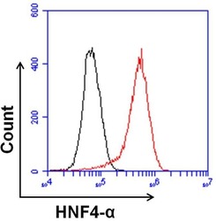
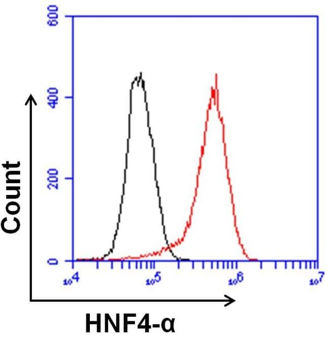
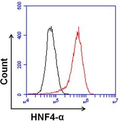
- Flow cytometry analysis of HNF4 alpha was done on HepG2 cells. Cells were fixed, permeabilized and stained with a HNF4 alpha mouse monoclonal antibody (Product # MA1-199, red histogram) or a mouse IgG2a isotype control (Product # MA1-10418, black histogram) at a dilution of 10 µg/mL. After incubation for 1 hour on ice, the cells were labeled with a Goat anti-Mouse IgG Secondary Antibody, DyLight 650 conjugate (Product # 84545) at a dilution of 1:50 for 1 hour on ice. A representative 10,000 cells were acquired and analyzed for each sample.
- Submitted by
- Invitrogen Antibodies (provider)
- Main image
- Experimental details
- Flow cytometry analysis of HNF4 alpha was done on HepG2 cells. Cells were fixed, permeabilized and stained with a HNF4 alpha mouse monoclonal antibody (Product # MA1-199, red histogram) or a mouse IgG2a isotype control (Product # MA1-10418, black histogram) at a dilution of 10 µg/mL. After incubation for 1 hour on ice, the cells were labeled with a Goat anti-Mouse IgG Secondary Antibody, DyLight 650 conjugate (Product # 84545) at a dilution of 1:50 for 1 hour on ice. A representative 10,000 cells were acquired and analyzed for each sample.

Supportive validation
- Submitted by
- Invitrogen Antibodies (provider)
- Main image
- Experimental details
- NULL
- Submitted by
- Invitrogen Antibodies (provider)
- Main image

- Experimental details
- NULL
- Submitted by
- Invitrogen Antibodies (provider)
- Main image

- Experimental details
- NULL
- Submitted by
- Invitrogen Antibodies (provider)
- Main image
- Experimental details
- NULL

- Submitted by
- Invitrogen Antibodies (provider)
- Main image
- Experimental details
- NULL
- Submitted by
- Invitrogen Antibodies (provider)
- Main image

- Experimental details
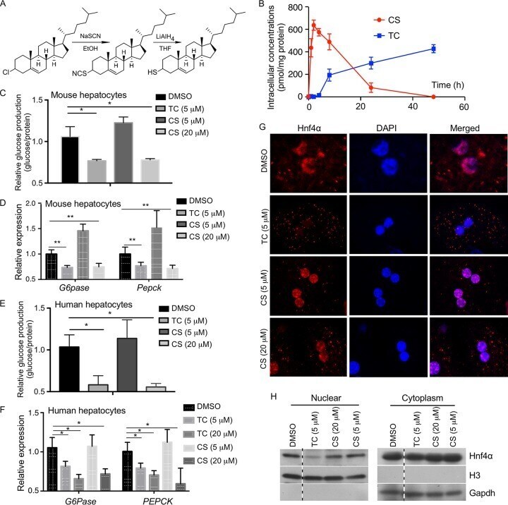
- FIG 7 Thiocholesterol (TC) shows an improved intracellular stability and better efficacy in inhibiting gluconeogenesis in primary hepatocytes. (A) Schematic depiction of the synthesis of TC from cholesteryl chloride. (B) Mouse primary hepatocytes isolated from 8-week-old male WT mice were treated with 5 muM TC or CS for up to 48 h, and the intracellular concentrations of TC and CS were measured by using UPLC-mass spectrometry. (C and D) Glucose production (C) and expression of gluconeogenic genes (D) in mouse primary hepatocytes treated with the indicated concentrations of drugs for 24 h. Cells were treated with 10 muM FSK for 2.5 h before the glucose production assay. The expression of each gene or glucose production was arbitrarily set as 1 in cells treated with DMSO. (E and F) The designs of the experiments were similar to those described for panels C and D, except that human primary hepatocytes were used. (G) The subcellular distribution of Hnf4alpha was visualized by immunofluorescence using an anti-Hnf4alpha antibody (red) in primary hepatocytes isolated from WT mice and treated with the indicated concentrations of drugs. DAPI (blue) was used for nuclear counterstaining. (H) The subcellular distribution of Hnf4alpha was measured by nuclear-cytosolic fractionation of the hepatocytes and Western blotting. Histone H3 and glyceraldehyde-3-phosphate dehydrogenase (Gapdh) are nuclear and cytoplasmic markers, respectively. All four lanes are from the