 Explore
 Validate
 Learn
Learn Western blot
 Immunocytochemistry
Antibody data
- Antibody Data
- Antigen structure
- References [10]
- Comments [0]
- Validations
- Immunocytochemistry [1]
- Immunohistochemistry [1]
- Chromatin Immunoprecipitation [1]
Submit
Validation data
Reference
Comment
Report error
- Product number
- HPA004712 - Provider product page

- Provider
- Atlas Antibodies
- Proper citation
- Atlas Antibodies Cat#HPA004712, RRID:AB_1079075
- Product name
- Anti-HNF4A
- Antibody type
- Polyclonal
- Description
- Polyclonal Antibody against Human HNF4A, Gene description: hepatocyte nuclear factor 4, alpha, Alternative Gene Names: HNF4, MODY, MODY1, NR2A1, TCF14, Validated applications: ChIP, ICC, IHC, WB, Uniprot ID: P41235, Storage: Store at +4°C for short term storage. Long time storage is recommended at -20°C.
- Reactivity
- Human
- Host
- Rabbit
- Conjugate
- Unconjugated
- Isotype
- IgG
- Vial size
- 100 µl
- Concentration
- 0.1 mg/ml
- Storage
- Store at +4°C for short term storage. Long time storage is recommended at -20°C.
- Handling
- The antibody solution should be gently mixed before use.
Submitted references Cell-permeated peptide P-T3H2 inhibits malignancy on hepatocellular carcinoma through stabilizing HNF4α protein
Extracellular vesicle-mediated protein delivery to the liver.
Phosphomannomutase 2 (PMM2) variants leading to hyperinsulinism-polycystic kidney disease are associated with early-onset inflammatory bowel disease and gastric antral foveolar hyperplasia.
Identification and validation of NFIA as a novel prognostic marker in renal cell carcinoma.
Phenotypical, functional and transcriptomic comparison of two modified methods of hepatocyte differentiation from human induced pluripotent stem cells
Serum transferrin as a biomarker of hepatocyte nuclear factor 4 alpha activity and hepatocyte function in liver diseases
Development of 3D Hepatic Constructs Within Polysaccharide-Based Scaffolds with Tunable Properties
Nuclear receptor HNF4α performs a tumor suppressor function in prostate cancer via its induction of p21-driven cellular senescence
Inhibition of the glucose transporter SGLT2 with dapagliflozin in pancreatic alpha cells triggers glucagon secretion
Global Gene Expression Responses to Low- or High-Dose Radiation in a Human Three-Dimensional Tissue Model
Wu S, Xiao M, Liu F, Hong H, Ding C, Zhang X, Xie W
Discover Oncology 2024;15(1)
Discover Oncology 2024;15(1)
Extracellular vesicle-mediated protein delivery to the liver.
Ilahibaks NF, Roefs MT, Brans MAD, Blok CS, de Jager SCA, Schiffelers RM, Vader P, Lei Z, Sluijter JPG
Journal of extracellular biology 2023 Sep;2(9):e97
Journal of extracellular biology 2023 Sep;2(9):e97
Phosphomannomutase 2 (PMM2) variants leading to hyperinsulinism-polycystic kidney disease are associated with early-onset inflammatory bowel disease and gastric antral foveolar hyperplasia.
Kiparissi F, Dastamani A, Palm L, Azabdaftari A, Campos L, Gaynor E, Grünewald S, Uhlig HH, Kleta R, Böckenhauer D, Jones KDJ
Human genetics 2023 May;142(5):697-704
Human genetics 2023 May;142(5):697-704
Identification and validation of NFIA as a novel prognostic marker in renal cell carcinoma.
de Alwis R, Schoch S, Islam M, Möller C, Ljungberg B, Axelson H
The journal of pathology. Clinical research 2023 Jul;9(4):261-272
The journal of pathology. Clinical research 2023 Jul;9(4):261-272
Phenotypical, functional and transcriptomic comparison of two modified methods of hepatocyte differentiation from human induced pluripotent stem cells
Li R, Zhao Y, Yourick J, Sprando R, Gao X
Biomedical Reports 2022;16(5)
Biomedical Reports 2022;16(5)
Serum transferrin as a biomarker of hepatocyte nuclear factor 4 alpha activity and hepatocyte function in liver diseases
Guldiken N, Argemi J, Gurbuz B, Atkinson S, Oliverius M, Fila P, Hamesch K, Bruns T, Cabezas J, Lozano J, Mann J, Cao S, Mathurin P, Shah V, Trautwein C, Thursz M, Bataller R, Strnad P
BMC Medicine 2021;19(1)
BMC Medicine 2021;19(1)
Development of 3D Hepatic Constructs Within Polysaccharide-Based Scaffolds with Tunable Properties
Labour M, Le Guilcher C, Aid-Launais R, El Samad N, Lanouar S, Simon-Yarza T, Letourneur D
International Journal of Molecular Sciences 2020;21(10):3644
International Journal of Molecular Sciences 2020;21(10):3644
Nuclear receptor HNF4α performs a tumor suppressor function in prostate cancer via its induction of p21-driven cellular senescence
Wang Z, Li Y, Wu D, Yu S, Wang Y, Leung Chan F
Oncogene 2019;39(7):1572-1589
Oncogene 2019;39(7):1572-1589
Inhibition of the glucose transporter SGLT2 with dapagliflozin in pancreatic alpha cells triggers glucagon secretion
Bonner C, Kerr-Conte J, Gmyr V, Queniat G, Moerman E, Thévenet J, Beaucamps C, Delalleau N, Popescu I, Malaisse W, Sener A, Deprez B, Abderrahmani A, Staels B, Pattou F
Nature Medicine 2015;21(5):512-517
Nature Medicine 2015;21(5):512-517
Global Gene Expression Responses to Low- or High-Dose Radiation in a Human Three-Dimensional Tissue Model
Mezentsev A, Amundson S
Radiation Research 2011;175(6):677
Radiation Research 2011;175(6):677
No comments: Submit comment
Supportive validation
- Submitted by
- Atlas Antibodies (provider)
- Main image
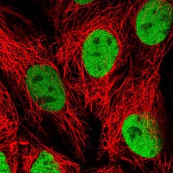
- Experimental details
- Immunofluorescent staining of human cell line CACO-2 shows localization to nucleoplasm.
- Sample type
- Human

Supportive validation
- Submitted by
- Atlas Antibodies (provider)
- Enhanced method
- Orthogonal validation
- Main image
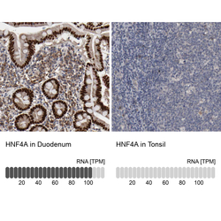
- Experimental details
- Immunohistochemistry analysis in human duodenum and tonsil tissues using HPA004712 antibody. Corresponding HNF4A RNA-seq data are presented for the same tissues.
- Sample type
- Human
- Protocol
- Protocol

Supportive validation
- Submitted by
- Atlas Antibodies (provider)
- Main image
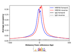
- Experimental details
- ChIP-Exo-Seq composite graph for Anti-HNF4A (HPA004712, Lot 000035854) tested in HepG2 cells. Strand-specific reads (blue: forward, red: reverse) and IgG controls (black: forward, grey: reverse) are plotted against the distance from a composite set of reference binding sites. The antibody exhibits robust target enrichment compared to a non-specific IgG control and precisely reveals its structural organization around the binding site. Data generated by Prof. B. F. Pugh´s Lab at Cornell University.
