Explore
 Validate
 Learn
Learn Western blot
 Immunocytochemistry
Antibody data
- Antibody Data
- Antigen structure
- References [2]
- Comments [0]
- Validations
- Immunocytochemistry [1]
Submit
Validation data
Reference
Comment
Report error
- Product number
- HPA004246 - Provider product page

- Provider
- Atlas Antibodies
- Proper citation
- Atlas Antibodies Cat#HPA004246, RRID:AB_1078151
- Product name
- Anti-PRKAG2
- Antibody type
- Polyclonal
- Description
- Polyclonal Antibody against Human PRKAG2, Gene description: protein kinase, AMP-activated, gamma 2 non-catalytic subunit, Alternative Gene Names: AAKG, AAKG2, CMH6, H91620p, WPWS, Validated applications: ICC, IHC, WB, Uniprot ID: Q9UGJ0, Storage: Store at +4°C for short term storage. Long time storage is recommended at -20°C.
- Reactivity
- Human, Mouse, Rat
- Host
- Rabbit
- Conjugate
- Unconjugated
- Isotype
- IgG
- Vial size
- 100 µl
- Concentration
- 0.1 mg/ml
- Storage
- Store at +4°C for short term storage. Long time storage is recommended at -20°C.
- Handling
- The antibody solution should be gently mixed before use.
Submitted references Subunit composition of AMPK trimers present in the cytokinetic apparatus: Implications for drug target identification
Generation of Digital Responses in Stress Sensors
Pinter K, Jefferson A, Czibik G, Watkins H, Redwood C
Cell Cycle 2014;11(5):917-921
Cell Cycle 2014;11(5):917-921
Generation of Digital Responses in Stress Sensors
Martiáñez T, Francès S, López J
Journal of Biological Chemistry 2009;284(36):23902-23911
Journal of Biological Chemistry 2009;284(36):23902-23911
No comments: Submit comment
Supportive validation
- Submitted by
- Atlas Antibodies (provider)
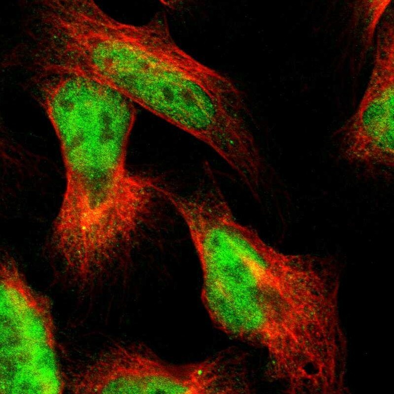
- Main image

- Experimental details
- Immunofluorescent staining of human cell line U-2 OS shows localization to nucleoplasm.
- Sample type
- Human