 Explore
 Validate
 Learn
Learn Immunocytochemistry
 Flow cytometry
Antibody data
- Antibody Data
- Antigen structure
- References [1]
- Comments [0]
- Validations
- Immunocytochemistry [1]
- Other assay [1]
Submit
Validation data
Reference
Comment
Report error
- Product number
- MA1-81793 - Provider product page

- Provider
- Invitrogen Antibodies
- Product name
- Glucocorticoid Receptor Monoclonal Antibody (5E4), FITC
- Antibody type
- Monoclonal
- Antigen
- Synthetic peptide
- Description
- Membrane permeabilization is required for flow cytometry applications. For FACS analysis, use 10 µL of the suggested working dilution to label 1x10^6 cells in 100 µL. Mouse anti Human Glucocorticoid Receptor antibody, clone 5E4 recognizes the human glucocorticoid receptor, also known as Nuclear receptor subfamily 3 group C member 1 (NR3C1), a 777 amino acid receptor bearing 3 distinct functional domains, an N-terminal modulating domain, a DNA binding domain and a C-terminal steroid binding domain.
- Reactivity
- Human
- Host
- Mouse
- Conjugate
- Green dye
- Isotype
- IgG
- Antibody clone number
- 5E4
- Vial size
- 100 µg
- Concentration
- 0.1 mg/mL
- Storage
- Store at 4°C short term. For long term storage, store at -20°C, avoiding freeze/thaw cycles. Store in the dark.
Submitted references Glucocorticoid Receptor Expression in Peripheral WBCs of Critically Ill Children.
Shibata AR, Troster EJ, Wong HR
Pediatric critical care medicine : a journal of the Society of Critical Care Medicine and the World Federation of Pediatric Intensive and Critical Care Societies 2015 Jun;16(5):e132-40
Pediatric critical care medicine : a journal of the Society of Critical Care Medicine and the World Federation of Pediatric Intensive and Critical Care Societies 2015 Jun;16(5):e132-40
No comments: Submit comment
Supportive validation
- Submitted by
- Invitrogen Antibodies (provider)
- Main image
- Experimental details
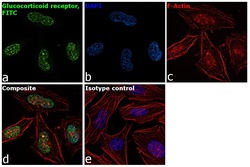
- Immunofluorescence analysis of Glucocorticoid Receptor was performed using 70% confluent log phase HeLa cells. The cells were fixed with 4% paraformaldehyde for 10 minutes, permeabilized with 0.1% Triton™ X-100 for 15 minutes, and blocked with 1% BSA for 1 hour at room temperature. The cells were labeled with Glucocorticoid Receptor, FITC Mouse Monoclonal Antibody (5E4) (Product # MA1-81793) at 5 µg/mL in 0.1% BSA and incubated at 4 degree Celsius overnight (Panel a: green). Nuclei (Panel b: blue) were stained with ProLong™ Diamond Antifade Mountant with DAPI (Product # P36962). F-actin (Panel c: red) was stained with Rhodamine Phalloidin (Product # R415, 1:300). Panel d represents the merged image showing nuclear localization. Panel e represents isotype control to assess background. The images were captured at 60X magnification.
- Conjugate
- Green dye

Supportive validation
- Submitted by
- Invitrogen Antibodies (provider)
- Main image

- Experimental details
- Figure 1. Glucocorticoid receptor (GCR) expression in CD4 lymphocytes, CD8 lymphocytes, monocytes (CD14), and neutrophils (CD66b) in patients with cardiovascular (CV) failure and without CV failure. GCR expression was lower in patients with CV failure when compared with patients without CV failure, p < 0.05 by rank-sum test. MFI = mean fluorescence intensity.
- Conjugate
- Green dye
