Explore
 Validate
 Learn
Learn Western blot
 Immunocytochemistry
Antibody data
- Antibody Data
- Antigen structure
- References [1]
- Comments [0]
- Validations
- Western blot [1]
Submit
Validation data
Reference
Comment
Report error
- Product number
- PB9232 - Provider product page

- Provider
- Boster Biological Technology
- Product name
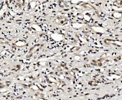
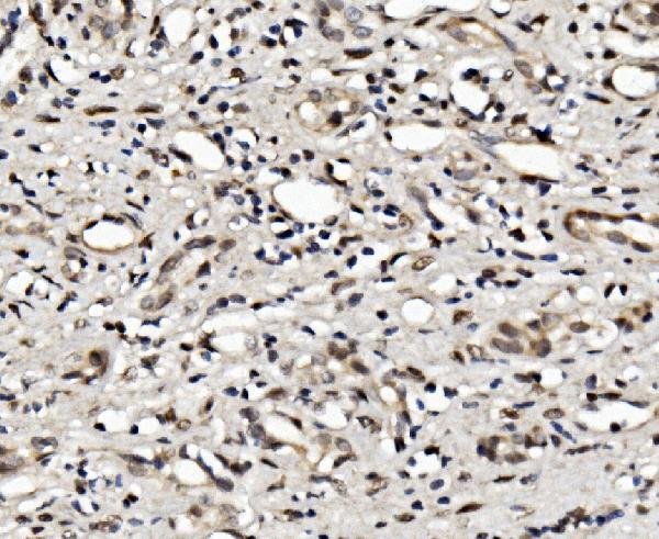
- Anti-Glucocorticoid Receptor/NR3C1 Antibody Picoband™
- Antibody type
- Polyclonal
- Description
- Polyclonal antibody for GLUCOCORTICOID RECEPTOR/NR3C1 detection. Host: Rabbit.Size: 100μg/vial. Tested applications: WB, IHC-P, ICC/IF, FCM. Reactive species: Human;Rat. GLUCOCORTICOID RECEPTOR/NR3C1 information: Molecular Weight: 85659 MW; Subcellular Localization: Cytoplasm . Mitochondrion. Nucleus . Cytoplasmic in the absence of ligand, nuclear after ligand- binding; Tissue Specificity: Widely expressed. In the heart, detected in left and right atria, left and right ventricles, aorta, apex, intraventricular septum, and atrioventricular node as well as whole adult and fetal heart.
- Reactivity
- Human, Rat
- Host
- Rabbit
- Vial size
- 100μg/vial
- Concentration
- Add 0.2ml of distilled water will yield a concentration of 500ug/ml.
- Storage
- At -20°C for one year. After reconstitution, at 4°C for one month. It can also be aliquoted and stored frozen at -20°C for a longer time. Avoid repeated freezing and thawing.
- Handling
- Add 0.2ml of distilled water will yield a concentration of 500ug/ml.
Submitted references CircDYM ameliorates CUMS mice depressive-like behavior and inhibits hippocampal neurons injury via miR-497a-5p/NR3C1 axis.
Li X, Sun X, Xie J, Wan H
Brain research 2022 Jul 15;1787:147911
Brain research 2022 Jul 15;1787:147911
No comments: Submit comment
Supportive validation
- Submitted by
- Boster Biological Technology (provider)
- Main image
- Experimental details
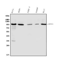
- Western blot analysis of NR3C1 using anti-NR3C1 antibody (PB9232). Electrophoresis was performed on a 5-20% SDS-PAGE gel at 70V (Stacking gel) / 90V (Resolving gel) for 2-3 hours. The sample well of each lane was loaded with 50ug of sample under reducing conditions. Lane 1: human HELA whole cell lysates, Lane 2: human A549 whole cell lysates, Lane 3: human PANC-1 whole cell lysates, Lane 4: human SW620 whole cell lysates, Lane 5: human Raji whole cell lysates. After Electrophoresis, proteins were transferred to a Nitrocellulose membrane at 150mA for 50-90 minutes. Blocked the membrane with 5% Non-fat Milk/ TBS for 1.5 hour at RT. The membrane was incubated with rabbit anti-NR3C1 antigen affinity purified polyclonal antibody (Catalog # PB9232) at 0.5 μg/mL overnight at 4°C, then washed with TBS-0.1%Tween 3 times with 5 minutes each and probed with a goat anti-rabbit IgG-HRP secondary antibody at a dilution of 1:5000 for 1.5 hour at RT. The signal is developed using an Enhanced Chemiluminescent detection (ECL) kit (Catalog # EK1002) with Tanon 5200 system. A specific band was detected for NR3C1 at approximately 100KD. The expected band size for NR3C1 is at 86KD.
- Additional image