Explore
 Validate
 Learn
Learn49-1026
antibody from Invitrogen Antibodies
Targeting: NR3C1
GR, GRL
 Western blot
 ELISA
ELISA Immunocytochemistry Immunoprecipitation Immunohistochemistry Chromatin Immunoprecipitation Other assay
Antibody data
- Antibody Data
- Antigen structure
- References [0]
- Comments [0]
- Validations
- Immunocytochemistry [2]
- Chromatin Immunoprecipitation [2]
- Other assay [1]
Submit
Validation data
Reference
Comment
Report error
- Product number
- 49-1026 - Provider product page

- Provider
- Invitrogen Antibodies
- Product name
- Glucocorticoid Receptor Monoclonal Antibody
- Antibody type
- Monoclonal
- Antigen
- Other
- Reactivity
- Human, Rat
- Host
- Mouse
- Isotype
- IgG
- Vial size
- 50 µg
- Concentration
- 1 mg/mL
- Storage
- -20° C, Avoid Freeze/Thaw Cycles
No comments: Submit comment
Supportive validation
- Submitted by
- Invitrogen Antibodies (provider)
- Main image
- Experimental details
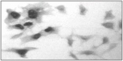
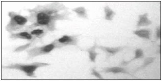
- COS-7 cells transiently overexpressing human GR were labeled with the monoclonal anti-hGR antibody followed by biotinylated second antibody and peroxidase-labeled avidin. The monoclonal antibody directed against GR (clone: m2F8) is used at 2.5 µg8260 mL.
- Submitted by
- Invitrogen Antibodies (provider)
- Main image
- Experimental details
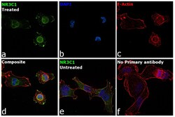
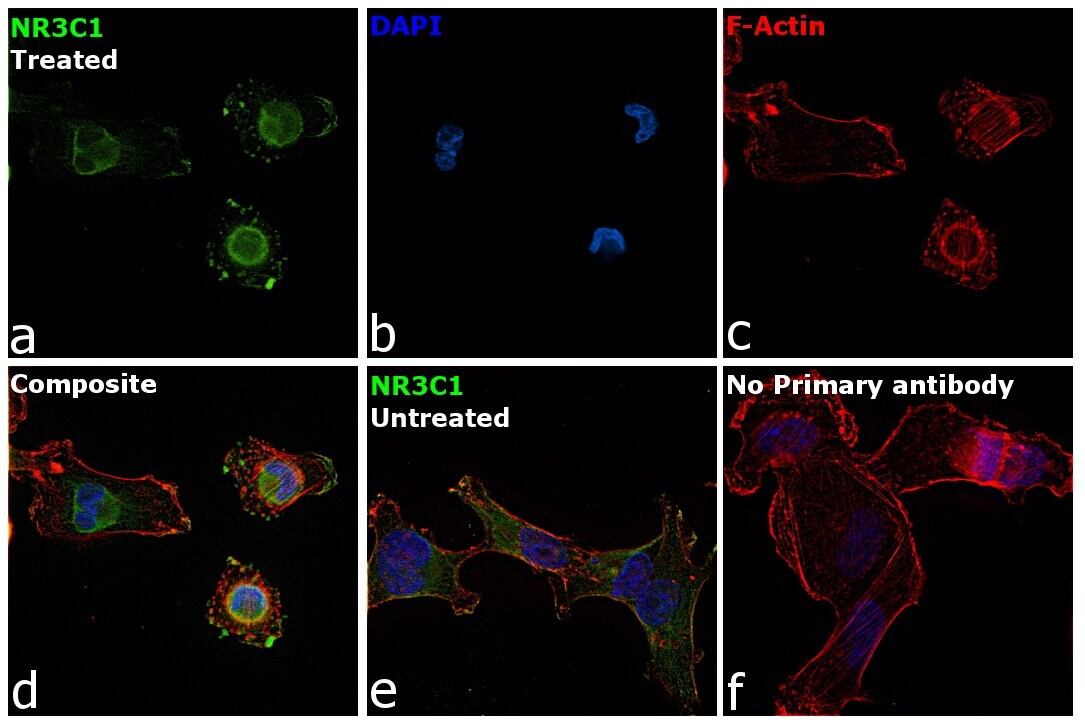
- Immunofluorescence analysis of Glucocorticoid Receptor (NR3C1) was performed using MDA-MB-231 cells (serum-starved) and MDA-MB-231 cells serum-starved for 24 hours, followed by 1 µM Dexamethasone treatment for 2 hours. The cells were fixed with 4% paraformaldehyde for 10 minutes, permeabilized with 0.1% Triton™ X-100 for 15 minutes, and blocked with 2% BSA for 45 minutes at room temperature. The cells were labeled with Glucocorticoid Receptor Monoclonal Antibody (Product # 49-1026) at 5 µg/mL in 0.1% BSA, incubated at 4 degree celsius overnight and then labeled with Donkey anti-Mouse IgG (H+L) Highly Cross-Adsorbed Secondary Antibody, Alexa Fluor Plus 488 (Product # A32766), (1:2000), for 45 minutes at room temperature (Panel a: Green) in MDA-MB-231 treated cells. Nuclei (Panel b:Blue) were stained with ProLong™ Diamond Antifade Mountant with DAPI (Product # P36962). F-actin (Panel c: Red) was stained with Rhodamine Phalloidin (Product # R415, 1:300). Panel d represents the merged image showing nuclear localization of NR3C1 protein in MDA-MB-231 treated cells. Panel e represents the merged image of MDA-MB-231 untreated cells, that shows cytoplasmic localization of NR3C1 protein. Panel f represents control cells with no primary antibody to assess background. The images were captured at 60X magnification.
Supportive validation
- Submitted by
- Invitrogen Antibodies (provider)
- Main image
- Experimental details
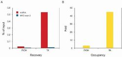
- ChIP assays were performed using HeLa cells, the anti-hGR monoclonal antibody (Product # 49-1026, clone: m2F8), and optimized PCR primer sets for qPCR. The cells were treated either with ethanol (EtOH) or triamcinolone acetonide (TA) for 4 hours prior to cell harvesting. Each ChIP assay used sheared chromatin from 3 million cells and 5 µg of the anti-hGR antibody. Recovery (%: ChIP⁄input; red bars) and occupancy (x fold: +ve⁄-ve; yellow bars)of human metallothionein promoter (hMTIIA) by GR, respectively, are shown. The recovery of human myoglobin exon 2 (hmyo ex2) by GR (%: ChIP⁄input; blue bars) was carried out as a negative control. Occupancy of the human metallothionein IIA promoter by GR is evident based on fluorescent qPCR analysis of immunoprecipitated DNA. Controls for IP and PCR specificity include primers for human myoglobin exon 2 (blue, negative control) and ethanol-treated cells.

- Submitted by
- Invitrogen Antibodies (provider)
- Main image
- Experimental details
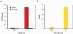
- ChIP assays were performed using HeLa cells, the anti-hGR monoclonal antibody (Product # 49-1026, clone: m2F8), and optimized PCR primer sets for qPCR. The cells were treated either with ethanol (EtOH) or triamcinolone acetonide (TA) for 4 hours prior to cell harvesting. Each ChIP assay used sheared chromatin from 3 million cells and 5 µg of the anti-hGR antibody. Recovery (%: ChIP⁄input; red bars) and occupancy (x fold: +ve⁄-ve; yellow bars)of human metallothionein promoter (hMTIIA) by GR, respectively, are shown. The recovery of human myoglobin exon 2 (hmyo ex2) by GR (%: ChIP⁄input; blue bars) was carried out as a negative control. Occupancy of the human metallothionein IIA promoter by GR is evident based on fluorescent qPCR analysis of immunoprecipitated DNA. Controls for IP and PCR specificity include primers for human myoglobin exon 2 (blue, negative control) and ethanol-treated cells.

Supportive validation
- Submitted by
- Invitrogen Antibodies (provider)
- Main image
- Experimental details
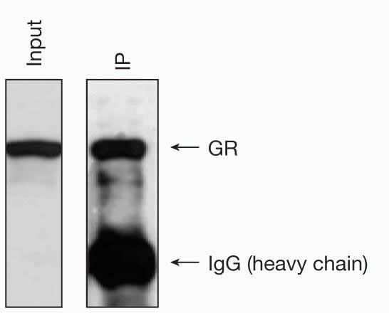
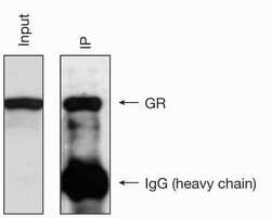
- Immunoprecipitation using the anti-hGR antibody. The immunoprecipitation (IP) of the glucocorticoid receptor from HeLa cell extracts is performed using the monoclonal antibody directed against GR (clone: m2F8) as follows: 5 µg of antibody per 5 million HeLa cells in 100 µl IP reaction solution. The IP is followed by western blot analysis with the same antibody.