Explore
 Validate
 Learn
Learn Western blot
 Immunoprecipitation
Antibody data
- Antibody Data
- Antigen structure
- References [2]
- Comments [0]
- Validations
- Immunoprecipitation [1]
- Immunohistochemistry [1]
- Other assay [3]
Submit
Validation data
Reference
Comment
Report error
- Product number
- PA5-21341 - Provider product page

- Provider
- Invitrogen Antibodies
- Product name
- Glucocorticoid Receptor Polyclonal Antibody
- Antibody type
- Polyclonal
- Antigen
- Recombinant full-length protein
- Description
- Recommended positive controls: HeLa, A431, Mouse heart, Rat heart. Predicted reactivity: Dog (88%), Cat (89%), Pig (83%), Sheep (89%), Bovine (90%), Guinea pig (85%). Store product as a concentrated solution. Centrifuge briefly prior to opening the vial.
- Reactivity
- Human, Mouse, Rat
- Host
- Rabbit
- Isotype
- IgG
- Vial size
- 100 μL
- Concentration
- 1 mg/mL
- Storage
- Store at 4°C short term. For long term storage, store at -20°C, avoiding freeze/thaw cycles.
Submitted references Long-Term Effects of Prenatal Severe Hypoxia on Central and Peripheral Components of the Glucocorticoid System in Rats.
Conditional deletion of glucocorticoid receptors in rat brain results in sex-specific deficits in fear and coping behaviors.
Vetrovoy O, Tyulkova E, Stratilov V, Baranova K, Nimiritsky P, Makarevich P, Rybnikova E
Developmental neuroscience 2020;42(2-4):145-158
Developmental neuroscience 2020;42(2-4):145-158
Conditional deletion of glucocorticoid receptors in rat brain results in sex-specific deficits in fear and coping behaviors.
Scheimann JR, Moloney RD, Mahbod P, Morano RL, Fitzgerald M, Hoskins O, Packard BA, Cotella EM, Hu YC, Herman JP
eLife 2019 Jul 22;8
eLife 2019 Jul 22;8
No comments: Submit comment
Supportive validation
- Submitted by
- Invitrogen Antibodies (provider)
- Main image

- Experimental details
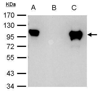
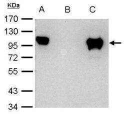
- Glucocorticoid receptor antibody immunoprecipitates Glucocorticoid receptor protein in IP experiments. IP Sample: 1,000 µg HeLa whole cell lysate/extract A. 40 µg HeLa whole cell lysate/extract B. Control with 2.5 µg of preimmune rabbit IgG C. Immunoprecipitation of Glucocorticoid receptor protein by 2.5 µg of Glucocorticoid receptor antibody (Product # PA5-21341) 7.5% SDS-PAGE The immunoprecipitated Glucocorticoid receptor protein was detected by SCMH1 antibody (Product # PA5-21341) diluted at 1:1,000.
Supportive validation
- Submitted by
- Invitrogen Antibodies (provider)
- Main image
- Experimental details
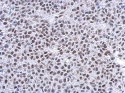
- Immunohistochemical analysis of paraffin-embedded Hela xenograft, using Glucocorticoid receptor (Product # PA5-21341) antibody at 1:500 dilution. Antigen Retrieval: EDTA based buffer, pH 8.0, 15 min.

Supportive validation
- Submitted by
- Invitrogen Antibodies (provider)
- Main image
- Experimental details
- Glucocorticoid receptor antibody immunoprecipitates Glucocorticoid receptor protein in IP experiments. IP Sample: 1,000 µg HeLa whole cell lysate/extract A. 40 µg HeLa whole cell lysate/extract B. Control with 2.5 µg of preimmune rabbit IgG C. Immunoprecipitation of Glucocorticoid receptor protein by 2.5 µg of Glucocorticoid receptor antibody (Product # PA5-21341) 7.5% SDS-PAGE The immunoprecipitated Glucocorticoid receptor protein was detected by SCMH1 antibody (Product # PA5-21341) diluted at 1:1,000.

- Submitted by
- Invitrogen Antibodies (provider)
- Main image
- Experimental details
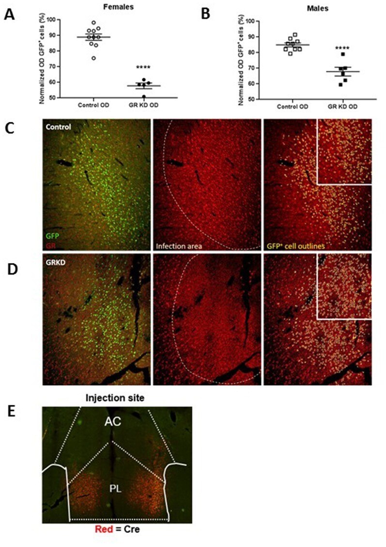
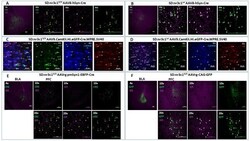
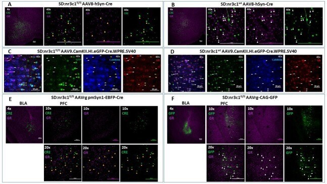
- Figure 2. Viral validation of conditional glucocorticoid receptor (GR) knockdown rat. ( A ) AAV8-hSyn-Cre administration to the basolateral amygdala (BLA) in SD:nr3c1 fl/fl rats (panel A1- site of injection, 4X). Note the absence of GR (purple) in Cre + neurons (green) (panels A2-4, 40x, yellow triangles). ( B ) AAV8-hSyn-Cre administration to the BLA in SD:nr3c1 wt rats (panel B1- site of injection, 4X). Note the presence of GR (purple) in Cre + neurons (green) (panels B2-4, 40x, white triangles). ( C ) AAV9.CamKII.HI.eGFP-Cre.WPRE.SV40 (CaMKIIalpha Cre) injected into SD:nr3c1 f/f rats results in decreased GR (C4 yellow arrows) in cells infected with virus as shown by GFP labeling (C2 yellow arrows) that are also CaMKIIalpha positive (C3 yellow arrows). CaMKIIalpha cells not infected with GFP, show endogenous GR expression (C1-4 white arrows). ( D ) CaMKIIalpha Cre injected into SD:nr3c1 wt rats (panel C2-4 40x white arrows) shows endogenous GR staining (D4) in cells infected with virus shown in GFP (D2) and CaMKIIalpha positive (D3). ( E ) AAVrg-pmSyn1-EBFP-Cre administration to the BLA in SD:nr3c1 fl/fl rats (panel E-1 site of injection, 4X) and retrograde trafficking of the virus to cell somas in the prefrontal cortex (PFC) (panel E2-4). Note absence of GR expression (purple) in Cre + neurons (green) (panel E5-7, 20x, yellow triangles). ( F ) AAVrg-CAG-GFP administration to the BLA in SD:nr3c1 fl/fl rats (panel F1- site of injection, 4X) and retrograde traffickin
- Submitted by
- Invitrogen Antibodies (provider)
- Main image
- Experimental details
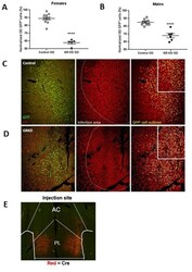
- 10.7554/eLife.44672.004 Figure 3. Verification of glucocorticoid receptor (GR) knockdown in the prelimbic division of the prefrontal cortex (PL-PFC). ( A ) Female SD:nr3c1 fl/fl rats injected with AAV9.CamKII.HI.eGFP-Cre.WPRE.SV40 show reduced GR expression in virus infected neurons (GFP + ) compared to controls (****p