Explore
 Validate
 Learn
Learn Western blot
 Immunocytochemistry
Antibody data
- Antibody Data
- Antigen structure
- References [1]
- Comments [0]
- Validations
- Immunocytochemistry [3]
- Immunohistochemistry [2]
Submit
Validation data
Reference
Comment
Report error
- Product number
- NB300-610 - Provider product page

- Provider
- Novus Biologicals
- Proper citation
- Novus Cat#NB300-610, RRID:AB_10002446
- Product name
- Rabbit Polyclonal GR/NR3C1 Antibody
- Antibody type
- Polyclonal
- Description
- Immunogen affinity purified.
- Reactivity
- Human, Mouse, Rat, Bovine, Rabbit, Sheep, Simian
- Host
- Rabbit
- Isotype
- IgG
- Vial size
- 50ug
- Concentration
- LYOPH
- Storage
- Store at -20C. Avoid freeze-thaw cycles.
Submitted references STAMP, a novel predicted factor assisting TIF2 actions in glucocorticoid receptor-mediated induction and repression.
He Y, Simons SS Jr
Molecular and cellular biology 2007 Feb;27(4):1467-85
Molecular and cellular biology 2007 Feb;27(4):1467-85
No comments: Submit comment
Supportive validation
- Submitted by
- Novus Biologicals (provider)
- Main image
- Experimental details
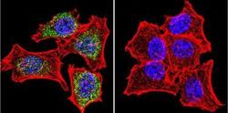
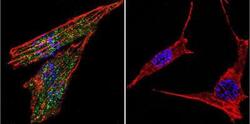
- Immunocytochemistry/Immunofluorescence: GR/NR3C1 Antibody [NB300-610] - Analysis of Glucocorticoid Receptor using Glucocorticoid Receptor Polyclonal Antibody shows staining in Hela Cells. Glucocorticoid Receptor (green), F-Actin staining with Phalloidin (red) and nuclei with DAPI (blue) is shown. Cells were grown on chamber slides and fixed with formaldehyde prior to staining. Cells were probed without (control) or with an antibody recognizing Glucocorticoid Receptor at a dilution of 1:20 over night at 4C, washed with PBS and incubated with a DyLight-488 conjugated.
- Submitted by
- Novus Biologicals (provider)
- Main image

- Experimental details
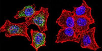
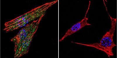
- Immunocytochemistry/Immunofluorescence: GR/NR3C1 Antibody [NB300-610] - Analysis of Glucocorticoid Receptor using Glucocorticoid Receptor Polyclonal Antibody shows staining in U251 Cells. Glucocorticoid Receptor (green), F-Actin staining with Phalloidin (red) and nuclei with DAPI (blue) is shown. Cells were grown on chamber slides and fixed with formaldehyde prior to staining. Cells were probed without (control) or with an antibody recognizing Glucocorticoid Receptor at a dilution of 1:20 over night at 4C, washed with PBS and incubated with a DyLight-488 conjugated.
- Submitted by
- Novus Biologicals (provider)
- Main image
- Experimental details
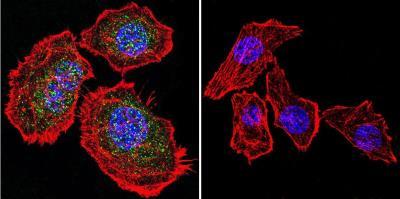
- Immunocytochemistry/Immunofluorescence: GR/NR3C1 Antibody [NB300-610] - Analysis of Glucocorticoid Receptor using Glucocorticoid Receptor Polyclonal Antibody shows staining in NIH-3T3 Cells. Glucocorticoid Receptor (green), F-Actin staining with Phalloidin (red) and nuclei with DAPI (blue) is shown. Cells were grown on chamber slides and fixed with formaldehyde prior to staining. Cells were probed without (control) or with an antibody recognizing Glucocorticoid Receptor at a dilution of 1:20 over night at 4C, washed with PBS and incubated with a DyLight-488 conjugated.
Supportive validation
- Submitted by
- Novus Biologicals (provider)
- Main image

- Experimental details
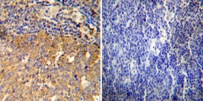
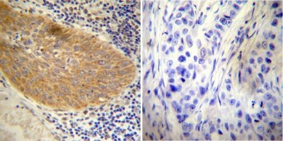
- Immunohistochemistry-Paraffin: GR/NR3C1 Antibody [NB300-610] - Both normal and cancer biopsies of deparaffinized human Cervical carcinoma tissue.
- Submitted by
- Novus Biologicals (provider)
- Main image

- Experimental details
- Immunohistochemistry-Paraffin: GR/NR3C1 Antibody [NB300-610] - Both normal and cancer biopsies of deparaffinized human Tonsil tissue.