Explore
 Validate
 Learn
Learn Western blot
Antibody data
- Antibody Data
- Antigen structure
- References [0]
- Comments [0]
- Validations
- Western blot [5]
- Immunocytochemistry [1]
- Immunohistochemistry [1]
- Flow cytometry [3]
Submit
Validation data
Reference
Comment
Report error
- Product number
- NBP2-29930 - Provider product page

- Provider
- Novus Biologicals
- Product name
- Rabbit Polyclonal SCAP Antibody
- Antibody type
- Polyclonal
- Description
- Protein A purified.
- Reactivity
- Human, Mouse, Rat
- Host
- Rabbit
- Isotype
- IgG
- Vial size
- 0.4 ml
- Concentration
- 0.77 mg/ml
- Storage
- Store at 4C short term. Aliquot and store at -20C long term. Avoid freeze-thaw cycles.
No comments: Submit comment
Supportive validation
- Submitted by
- Novus Biologicals (provider)
- Main image
- Experimental details
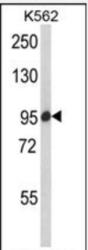
- Western Blot: SCAP Antibody [NBP2-29930] - K562 cell line lysates (35ug/lane). SCAP (arrow) was detected using the purified Pab.

- Submitted by
- Novus Biologicals (provider)
- Main image

- Experimental details
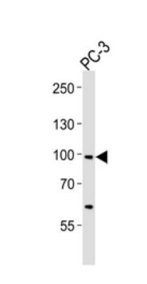
- Western Blot: SCAP Antibody [NBP2-29930] - PC-3 cell line
- Submitted by
- Novus Biologicals (provider)
- Main image

- Experimental details
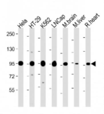
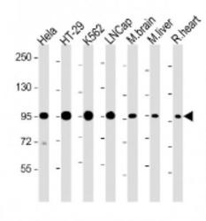
- Western Blot: SCAP Antibody [NBP2-29930] - Anti-SCAP Antibody (Center) at 1:2000 dilution Lane 1: Hela whole cell lysate Lane 2: HT-29 whole cell lysate Lane 3: K562 whole cell lysate Lane 4: LNCap whole cell lysate Lane 5: Mouse brain lysate Lane 6: Mouse liver lysate Lane 7: Rat heart lysate Lysates/proteins at 20 ug per lane. Secondary Goat Anti-Rabbit IgG, (H+L), Peroxidase conjugated at 1/10000 dilution. Predicted band size : 140, 98, 96 kDa Blocking/Dilution buffer: 5% NFDM/TBST.
- Submitted by
- Novus Biologicals (provider)
- Main image
- Experimental details
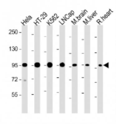
- Western Blot: SCAP Antibody [NBP2-29930] - Anti-SCAP Antibody (Center) at 1:2000 dilution Lane 1: Hela whole cell lysate Lane 2: HT-29 whole cell lysate Lane 3: K562 whole cell lysate Lane 4: LNCap whole cell lysate Lane 5: Mouse brain lysate Lane 6: Mouse liver lysate Lane 7: Rat heart lysate Lysates/proteins at 20 ug per lane. Secondary Goat Anti-Rabbit IgG, (H+L), Peroxidase conjugated at 1/10000 dilution. Predicted band size : 140, 98, 96 kDa Blocking/Dilution buffer: 5% NFDM/TBST.
- Submitted by
- Novus Biologicals (provider)
- Main image

- Experimental details
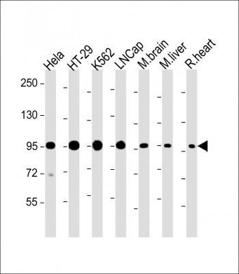
- Western Blot: SCAP Antibody (RB22508) [NBP2-29930] - All lanes : Anti-SCAP Antibody (Center). Lane 1: Hela whole cell lysate; Lane 2: HT-29 whole cell lysate; Lane 3: K562 whole cell lysate; Lane 4: LNCap whole cell lysate; Lane 5: Mouse brain lysate; Lane 6: Mouse liver lysate; Lane 7: Rat heart lysate. Lysates/proteins at 20 ug per lane. Secondary Goat Anti-Rabbit IgG, (H+L), Peroxidase conjugated. Predicted band size : 140, 98, 96 kDa. Blocking/Dilution buffer: 5% NFDM/TBST.
Supportive validation
- Submitted by
- Novus Biologicals (provider)
- Main image
- Experimental details
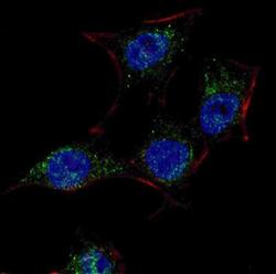
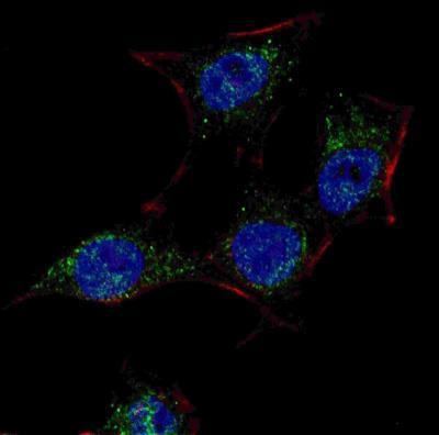
- Immunocytochemistry/Immunofluorescence: SCAP Antibody [NBP2-29930] - Fluorescent confocal image of HeLa cells stained with SCAP (Center) antibody. HeLa cells were fixed with 4% PFA (20 min), permeabilized with Triton X-100 (0.2%, 30 min). Cells were then incubated with AP8568c SCAP (Center) primary antibody (1:100, 2 h at room temperature). For secondary antibody, Alexa Fluor 488 conjugated donkey anti-rabbit antibody (green) was used (1:1000, 1h). Nuclei were counterstained with Hoechst 33342 (blue) (10 ug/ml, 5 min).
Supportive validation
- Submitted by
- Novus Biologicals (provider)
- Main image
- Experimental details
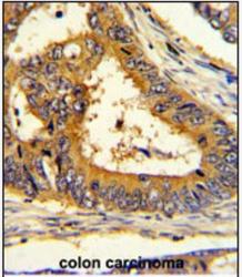
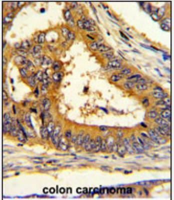
- Immunohistochemistry-Paraffin: SCAP Antibody [NBP2-29930] - Formalin-fixed and paraffin-embedded human colon carcinoma with (Center), which was peroxidase-conjugated to the secondary antibody, followed by DAB staining. This data demonstrates the use of this antibody for immunohistochemistry; clinical relevance has not been evaluated.
Supportive validation
- Submitted by
- Novus Biologicals (provider)
- Main image

- Experimental details
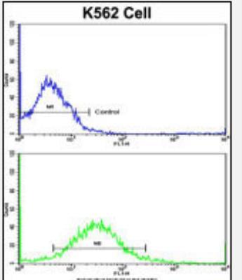
- Flow Cytometry: SCAP Antibody [NBP2-29930] - K562 cells using (Center)(bottom histogram) compared to a negative control cell (top histogram). FITC-conjugated goat-anti-rabbit secondary antibodies were used for the analysis.
- Submitted by
- Novus Biologicals (provider)
- Main image
- Experimental details
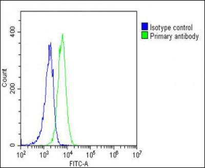
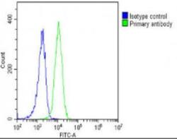
- Flow Cytometry: SCAP Antibody [NBP2-29930] - Overlay histogram showing K562 cells stained with AP8568c(green line). The cells were fixed with 2% paraformaldehyde (10 min) and then permeabilized with 90% methanol for 10 min. The cells were then icubated in 2% bovine serum albumin to block non-specific protein-protein interactions followed by the antibody (AP8568c, 1:25 dilution) for 60 min at 37 C. The secondary antibody used was Goat-Anti-Rabbit IgG, DyLight (R) 488 Conjugated Highly Cross-Adsorbed(OH191631) at 1/200 dilution for 40 min at 37 C. Isotype control antibody (blue line) was rabbit IgG1 (1ug/1x10^6 cells) used under the same conditions. Acquisition of >10, 000 events was performed.

- Submitted by
- Novus Biologicals (provider)
- Main image
- Experimental details
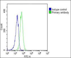
- Flow Cytometry: SCAP Antibody (RB22508) [NBP2-29930] - Overlay histogram showing K562 cells stained with NBP2-29930 (green line). The cells were fixed with 2% paraformaldehyde (10 min) and then permeabilized with 90% methanol for 10 min. The cells were then icubated in 2% bovine serum albumin to block non-specific protein-protein interactions followed by the antibody for 60 min at 37ºC. The secondary antibody used was Goat-Anti-Rabbit IgG, DyLight® 488 Conjugated Highly Cross-Adsorbed for 40 min at 37ºC. Isotype control antibody (blue line) was rabbit IgG1 (1ug/1x10^6 cells) used under the same conditions. Acquisition of >10, 000 events was performed.