Explore
 Validate
 Learn
LearnGTX15763
antibody from GeneTex
Targeting: CXCL8
3-10C, AMCF-I, b-ENAP, GCP-1, GCP1, IL-8, IL8, K60, LECT, LUCT, LYNAP, MDNCF, MONAP, NAF, NAP-1, NAP1, SCYB8, TSG-1
 Western blot
 ELISA
ELISAAntibody data
- Antibody Data
- Antigen structure
- References [0]
- Comments [0]
- Validations
- ELISA [1]
- Immunocytochemistry [1]
- Flow cytometry [1]
Submit
Validation data
Reference
Comment
Report error
- Product number
- GTX15763 - Provider product page

- Provider
- GeneTex
- Product name
- CXCL8 / IL8 antibody [3IL8-H10]
- Antibody type
- Monoclonal
- Reactivity
- Human
- Host
- Mouse
No comments: Submit comment
Supportive validation
- Submitted by
- GeneTex (provider)
- Main image
- Experimental details
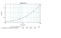
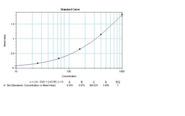
- Sandwich ELISA analysis of human IL-8 was performed using a Human IL-8 Colorimetric ELISA kit by loading 50 ul per well of Human IL-8 Recombinant Protein in dodecuplicate at 1000, 400, 160, 64, 25.6, and 0 pg/ml across a 3 ug/ml mouse anti-Human IL-8 (M801) pre-coated plate and incubating for 1 hour at room temperature. The plate was washed, and then incubated with 50 ul per well of a biotinylated mouse anti-human IL-8 monoclonal antibody in duplicate at 0.25 ug/ml for 1 hour at room temperature. The plate was washed and incubated with 100 ul per well of Streptavidin-HRP in all test wells at a 1:4,000 dilution for 30 minutes at room temperature. Detection was performed using 1-Step Ultra TMB substrate for 30 minutes at room temperature in the dark. The plate was then stopped with 0.16M sulfuric acid. Absorbances were read on a spectrophotometer at 450-550 nm.
Supportive validation
- Submitted by
- GeneTex (provider)
- Main image
- Experimental details
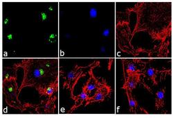
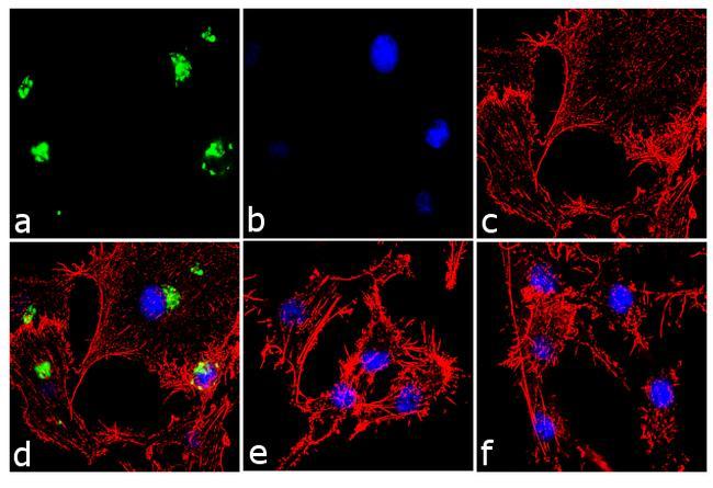
- Immunofluorescence analysis of IL-8 was performed using 70% confluent log phase U-87 MG cells treated 1 uM thapsigargin for 24 hours. The cells were fixed with 4% paraformaldehyde for 10 minutes, permeabilized with 0.1% Triton? X-100 for 10 minutes, and blocked with 1% BSA for 1 hour at room temperature. The cells were labeled with IL-8 Mouse Monoclonal Antibody (M801) at 2?g/ml in 0.1% BSA and incubated for 3 hours at room temperature and then labeled with Goat anti-Mouse IgG (H+L) Superclonal? Secondary Antibody, Alexa Fluor? 488 conj?gate at a dilution of 1:2000 for 45 minutes at room temperature (Panel a: green). Nuclei (Panel b: blue) were stained with SlowFade? Gold Antifade Mountant with DAPI. F-actin (Panel c: red) was stained with Alexa Fluor? 555 Rhodamine Phalloidin. Panel d represents the merged image showing cytoplasmic localization. Panel e is untreated cell with no signal. Panel f represents control cells with no primary antibody to assess background. The images were captured at 60X magnification..
Supportive validation
- Submitted by
- GeneTex (provider)
- Main image
- Experimental details
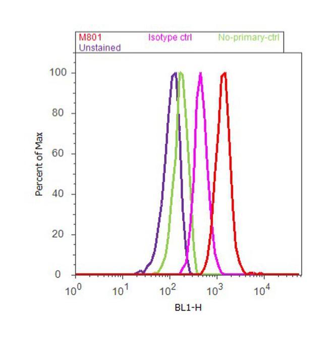
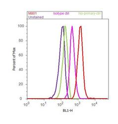
- Flow cytometry analysis of IL-8 was done on U-87 MG cells. Cells were fixed with 70% ethanol for 10 minutes, permeabilized with 0.25% Triton? X-100 for 20 minutes, and blocked with 5% BSA for 30 minutes at room temperature. Cells were labeled with IL-8 Mouse Monoclonal Antibody (M801, red histogram) or with mouse isotype control (pink histogram) at 3-5 ?g/million cells in 2.5% BSA. After incubation at room temperature for 2 hours, the cells were labeled with Alexa Fluor? 488 Rabbit Anti-Mouse Secondary Antibody at a dilution of 1:400 for 30 minutes at room temperature. The representative 10,000 cells were acquired and analyzed for each sample using an Attune? Acoustic Focusing Cytometer. The purple histogram represents unstained control cells and the green histogram represents no-primary-antibody control.