Explore
 Validate
 Learn
LearnMA5-23697
antibody from Invitrogen Antibodies
Targeting: CXCL8
3-10C, AMCF-I, b-ENAP, GCP-1, GCP1, IL-8, IL8, K60, LECT, LUCT, LYNAP, MDNCF, MONAP, NAF, NAP-1, NAP1, SCYB8, TSG-1
 Western blot
 ELISA
ELISA Immunocytochemistry
Antibody data
- Antibody Data
- Antigen structure
- References [2]
- Comments [0]
- Validations
- Immunocytochemistry [2]
- Other assay [1]
Submit
Validation data
Reference
Comment
Report error
- Product number
- MA5-23697 - Provider product page

- Provider
- Invitrogen Antibodies
- Product name
- IL-8 (CXCL8) Monoclonal Antibody (6217)
- Antibody type
- Monoclonal
- Antigen
- Recombinant full-length protein
- Description
- In Western blots, this antibody shows 100% cross-reactivity with recombinant porcine CXCL8/IL-8 and no cross-reactivity with rrCXCL3/CINC-2 alpha. Reconstitute at 0.5 mg/mL in sterile PBS. Endoxin level is
- Reactivity
- Human
- Host
- Mouse
- Isotype
- IgG
- Antibody clone number
- 6217
- Vial size
- 500 μg
- Concentration
- 0.5 mg/mL
- Storage
- -20°C, Avoid Freeze/Thaw Cycles
Submitted references Tumour cell apoptosis modulates the colorectal cancer immune microenvironment via interleukin-8-dependent neutrophil recruitment.
FABP4 deactivates NF-κB-IL1α pathway by ubiquitinating ATPB in tumor-associated macrophages and promotes neuroblastoma progression.
Schimek V, Strasser K, Beer A, Göber S, Walterskirchen N, Brostjan C, Müller C, Bachleitner-Hofmann T, Bergmann M, Dolznig H, Oehler R
Cell death & disease 2022 Feb 4;13(2):113
Cell death & disease 2022 Feb 4;13(2):113
FABP4 deactivates NF-κB-IL1α pathway by ubiquitinating ATPB in tumor-associated macrophages and promotes neuroblastoma progression.
Miao L, Zhuo Z, Tang J, Huang X, Liu J, Wang HY, Xia H, He J
Clinical and translational medicine 2021 Apr;11(4):e395
Clinical and translational medicine 2021 Apr;11(4):e395
No comments: Submit comment
Supportive validation
- Submitted by
- Invitrogen Antibodies (provider)
- Main image

- Experimental details
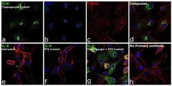
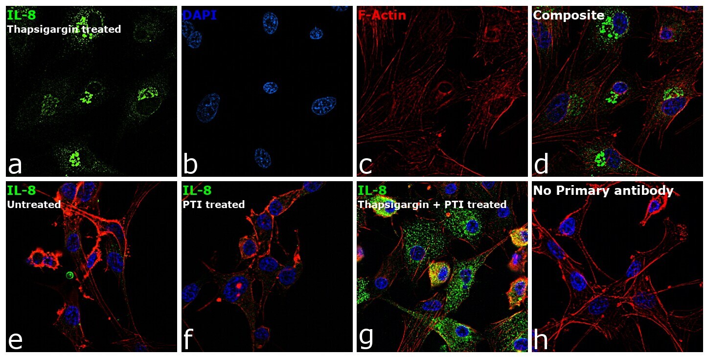
- Immunofluorescence analysis of IL-8 (CXCL8) was performed using 70% confluent log phase U-87 MG cells treated with 1uM of Thapsigargin for 24 hours. The cells were fixed with 4% paraformaldehyde for 10 minutes, permeabilized with 0.1% Triton™ X-100 for 15 minutes, and blocked with 2% BSA for 45 minutes at room temperature. The cells were labeled with IL-8 (CXCL8) Monoclonal Antibody (6217) (Product # MA5-23697) at 8 µg/mL in 0.1% BSA, incubated at 4 degree celsius overnight and then labeled with Goat anti-Mouse IgG (H+L) Highly Cross-Adsorbed Secondary Antibody, Alexa Fluor Plus 488 (Product # A32723), (1:2000), for 45 minutes at room temperature (Panel a: Green). Nuclei (Panel b:Blue) were stained with ProLong™ Diamond Antifade Mountant with DAPI (Product # P36962). F-actin (Panel c: Red) was stained with Rhodamine Phalloidin (Product # R415, 1:300). Panel d represents the merged image showing cytoplasmic(Golgi complex like pattern) localization. Panel e represents untreated cells . Panel f represents cells treated with PTI . Panel g represents cells treated with Thapsigargin and PTI. Panel h represents control cells with no primary antibody to assess background. The images were captured at 60X magnification.

- Submitted by
- Invitrogen Antibodies (provider)
- Main image
- Experimental details
- Immunofluorescence analysis of IL-8 (CXCL8) was performed using 70% confluent log phase U-87 MG cells treated with 1uM of Thapsigargin for 24 hours. The cells were fixed with 4% paraformaldehyde for 10 minutes, permeabilized with 0.1% Triton™ X-100 for 15 minutes, and blocked with 2% BSA for 45 minutes at room temperature. The cells were labeled with IL-8 (CXCL8) Monoclonal Antibody (6217) (Product # MA5-23697) at 8 µg/mL in 0.1% BSA, incubated at 4 degree celsius overnight and then labeled with Goat anti-Mouse IgG (H+L) Highly Cross-Adsorbed Secondary Antibody, Alexa Fluor Plus 488 (Product # A32723), (1:2000), for 45 minutes at room temperature (Panel a: Green). Nuclei (Panel b:Blue) were stained with ProLong™ Diamond Antifade Mountant with DAPI (Product # P36962). F-actin (Panel c: Red) was stained with Rhodamine Phalloidin (Product # R415, 1:300). Panel d represents the merged image showing cytoplasmic(Golgi complex like pattern) localization. Panel e represents untreated cells . Panel f represents cells treated with PTI . Panel g represents cells treated with Thapsigargin and PTI. Panel h represents control cells with no primary antibody to assess background. The images were captured at 60X magnification.
Supportive validation
- Submitted by
- Invitrogen Antibodies (provider)
- Main image
- Experimental details
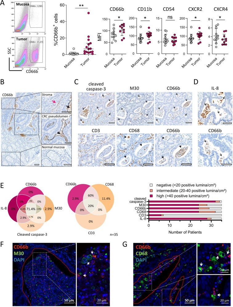
- Fig. 4 Neutrophils co-localise with apoptotic tumour cells and tumour-associated macrophages in CRC pseudolumina. A Flow cytometric analysis of dissociated CRC tissues and matched normal mucosae of treatment-naive CRC patients. For gating strategy, see Fig. S7A . Graphs present median and 95% CI (%CD66b+ cells, n = 15) or mean +- SD (surface marker MFIs, n = 10-12). * P < 0.05, ** P < 0.01, ns not significant, as calculated by Wilcoxon signed-rank test (%CD66b+ cells) or two-tailed paired t -tests (surface marker MFIs). B Representative CD66b immunohistochemistry (IHC) of a CRC tissue. Red and black arrows indicate stromal and intraluminal neutrophils, respectively, while dotted lines illustrate CRC pseudolumina. Scale bars, 100 um. C , D Consecutive IHC sections of representative CRC specimens stained for M30, cleaved caspase-3 and CD66b ( C upper panel), CD3, CD68 and CD66b ( C lower panel) and IL-8 and CD66b ( D ). Arrows indicate positive staining in CRC pseudolumina. Scale bars, 100 um. E Quantification of cleaved caspase-3-expressing, M30-expressing, IL-8-expressing, CD66b-expressing, CD68-expressing and CD3-expressing CRC pseudolumina in IHC sections of 35 CRC patients. Patients with at least 20 IHC-positive pseudolumina/cm 2 tissue area were considered positive in the presented Venn diagrams. F CD66b and M30 immunofluorescence staining of a CRC pseudolumen. Arrows indicate close contact between CD66b-positive neutrophils and M30-positive apoptotic tumour cells. Images