Explore
 Validate
 Learn
Learn17-8088-41
antibody from Invitrogen Antibodies
Targeting: CXCL8
3-10C, AMCF-I, b-ENAP, GCP-1, GCP1, IL-8, IL8, K60, LECT, LUCT, LYNAP, MDNCF, MONAP, NAF, NAP-1, NAP1, SCYB8, TSG-1
 Flow cytometry
Antibody data
- Antibody Data
- Antigen structure
- References [3]
- Comments [0]
- Validations
- Flow cytometry [1]
Submit
Validation data
Reference
Comment
Report error
- Product number
- 17-8088-41 - Provider product page

- Provider
- Invitrogen Antibodies
- Product name
- Anti-IL-8 (1-77) (CXCL8) Monoclonal Antibody (8CH), APC, eBioscience™
- Antibody type
- Monoclonal
- Antigen
- Other
- Description
- Description: This 8CH monoclonal antibody reacts with human IL-8 (CXCL8), a pro-inflammatory CXC chemokine. It is synthesized as a 99 amino acid precursor protein that is further processed into one of four isoforms, with the most common being 72 or 77 amino acids in length. IL-8(77) is secreted primarily by endothelial cells and is thought to be a less potent neutrophil activator than the other forms. It is present at high levels during fetal development, where it mediates angiogenesis rather than inflammation. The predominant form present in adults is IL-8(72), which is expressed by monocytes, macrophages, epithelial cells, and fibroblasts in response to inflammatory stimuli, environmental stress, and steroid hormones. IL-8(72) is essential for the activation and recruitment of neutrophils to sites of inflammation, and has also been found to influence T cell migration. Signaling occurs through the G-protein coupled receptors CXCR1 or CXCR2. IL-8 transcripts are often upregulated in tumors, and it is associated with tumor angiogenesis and metastasis. Applications Reported: This 8CH antibody has been reported for use in intracellular staining followed by flow cytometric analysis. Applications Tested: This 8CH antibody has been pre-titrated and tested by intracellular staining followed by flow cytometric analysis. This can be used at 5 µL (0.015 µg) per test. A test is defined as the amount (µg) of antibody that will stain a cell sample in a final volume of 100 µL. Cell number should be determined empirically but can range from 10^5 to 10^8 cells/test. Excitation: 633-647 nm; Emission: 660 nm; Laser: Red Laser. Filtration: 0.2 µm post-manufacturing filtered.
- Reactivity
- Human
- Host
- Mouse
- Isotype
- IgG
- Antibody clone number
- 8CH
- Vial size
- 25 Tests
- Concentration
- 5 µL/Test
- Storage
- 4° C, store in dark, DO NOT FREEZE!
Submitted references Proteotranscriptomics Reveal Signaling Networks in the Ovarian Cancer Microenvironment.
A transcriptome-based global map of signaling pathways in the ovarian cancer microenvironment associated with clinical outcome.
The Processed Amino-Terminal Fragment of Human TLR7 Acts as a Chaperone To Direct Human TLR7 into Endosomes.
Worzfeld T, Finkernagel F, Reinartz S, Konzer A, Adhikary T, Nist A, Stiewe T, Wagner U, Looso M, Graumann J, Müller R
Molecular & cellular proteomics : MCP 2018 Feb;17(2):270-289
Molecular & cellular proteomics : MCP 2018 Feb;17(2):270-289
A transcriptome-based global map of signaling pathways in the ovarian cancer microenvironment associated with clinical outcome.
Reinartz S, Finkernagel F, Adhikary T, Rohnalter V, Schumann T, Schober Y, Nockher WA, Nist A, Stiewe T, Jansen JM, Wagner U, Müller-Brüsselbach S, Müller R
Genome biology 2016 May 23;17(1):108
Genome biology 2016 May 23;17(1):108
The Processed Amino-Terminal Fragment of Human TLR7 Acts as a Chaperone To Direct Human TLR7 into Endosomes.
Hipp MM, Shepherd D, Booth S, Waithe D, Reis e Sousa C, Cerundolo V
Journal of immunology (Baltimore, Md. : 1950) 2015 Jun 1;194(11):5417-25
Journal of immunology (Baltimore, Md. : 1950) 2015 Jun 1;194(11):5417-25
No comments: Submit comment
Supportive validation
- Submitted by
- Invitrogen Antibodies (provider)
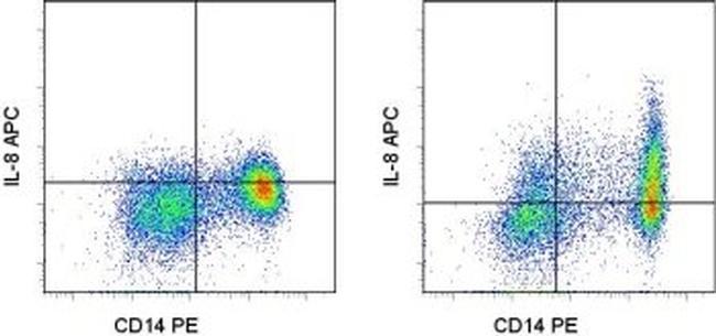
- Main image
- Experimental details
- Normal human peripheral blood cells unstimulated (left) or stimulated with LPS in the presence of Protein Transport Inhibitor Cocktail (Product # 00-4980-03), stained with Anti-Human CD14 PE (Product # 12-0149-42) followed by fixation and intracellular staining with and Anti-Human IL-8 APC. Cells in the monocyte gate were used for analysis.