Explore
 Validate
 Learn
Learn14-7189-80
antibody from Invitrogen Antibodies
Targeting: CXCL8
3-10C, AMCF-I, b-ENAP, GCP-1, GCP1, IL-8, IL8, K60, LECT, LUCT, LYNAP, MDNCF, MONAP, NAF, NAP-1, NAP1, SCYB8, TSG-1
 Western blot
Antibody data
- Antibody Data
- Antigen structure
- References [0]
- Comments [0]
- Validations
- Western blot [2]
- Immunocytochemistry [1]
Submit
Validation data
Reference
Comment
Report error
- Product number
- 14-7189-80 - Provider product page

- Provider
- Invitrogen Antibodies
- Product name
- IL-8 (1-77) (CXCL8) Monoclonal Antibody (N11), eBioscience™
- Antibody type
- Monoclonal
- Antigen
- Other
- Description
- Description: The monoclonal antibody N11 reacts specifically with the 77 amino acid form of IL-8 (IL-877, IL-8(1-77), CXCL877) and does not react with the more common 72 amino acid form. Frequently referred to as "endothelial" IL-8, the 77 amino acid form is secreted in cultured leukocytes. The 72 amino acid form is the result of efficient cleavage of an N-terminal pentamer from IL-877 after secretion. IL-8 is a C-X-C chemokine with potency in neutrophil chemotaxis and activation. IL-877 has decreased chemotactic activity when compared to IL-872, but has other specific pro-apoptotic and tumor cell chemotactic activities. IL-877 has recently been shown to be the major isoform present in premature neonates due to deficiency in serum protease activity that is not seen in term neonates.
- Antibody clone number
- N11
- Concentration
- 0.5 mg/mL
No comments: Submit comment
Supportive validation
- Submitted by
- Invitrogen Antibodies (provider)
- Main image

- Experimental details
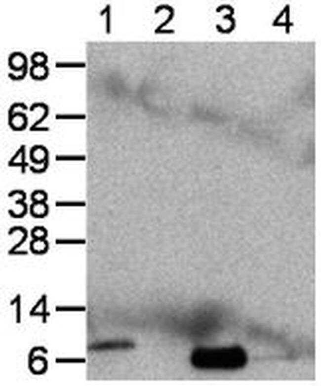
- Immunoblot of human peripheral blood mononuclear cells either left untreated or treated with LPS and Brefeldin A for 4 hours. Lane 1, non-reduced lysate prepared from stimulated cells; lane 2, non-reduced lysate of unstimulated cells; lane 3, reduced lysate of stimulated cells; lane 4, reduced lysate prepared from unstimulated cells. Bands were visualized using Anti-Mouse IgG HRP.
- Submitted by
- Invitrogen Antibodies (provider)
- Main image
- Experimental details
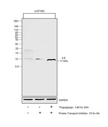
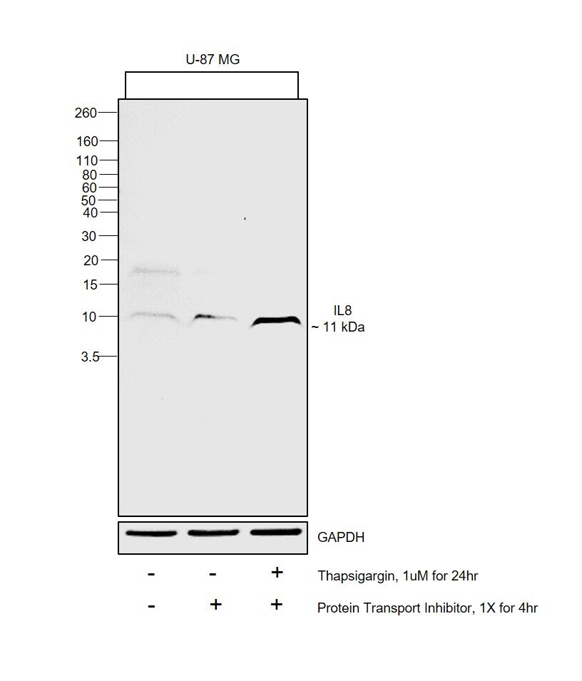
- Western blot was performed using Anti-IL-8 (1-77) (CXCL8) Monoclonal Antibody (N11), eBioscience™(Product # 14-7189-82) and a 11kDa band corresponding to IL-8 (1-77) (CXCL8) was observed in U-87 MG up on treatment with Thapsigargin and PTI. Whole cell extracts (60 µg lysate) of U-87 MG (Lane 1), U-87 MG treated with PTI (1X for 4hr) (Lane 2), U-87 MG treated with Thapsigargin (1uM for 24hr) followed by PTI (1X for 4hr) (Lane 3) were electrophoresed using Novex™ 16% Tricine Protein Gel (Product # EC6695BOX). Resolved proteins were then transferred onto a Nitrocellulose membrane (Product # IB23001) by iBlot® 2 Dry Blotting System (Product # IB21001). The blot was probed with the primary antibody (1 µg/mL) and detected by chemiluminescence with Goat anti-Mouse IgG (H+L) Superclonal™ Recombinant Secondary Antibody, HRP (Product # A28177, 1:4000 dilution) using the iBright FL 1000 (Product # A32752). Chemiluminescent detection was performed using SuperSignal™ West Atto Ultimate Sensitivity Substrate (Product # A38556).
Supportive validation
- Submitted by
- Invitrogen Antibodies (provider)
- Main image
- Experimental details
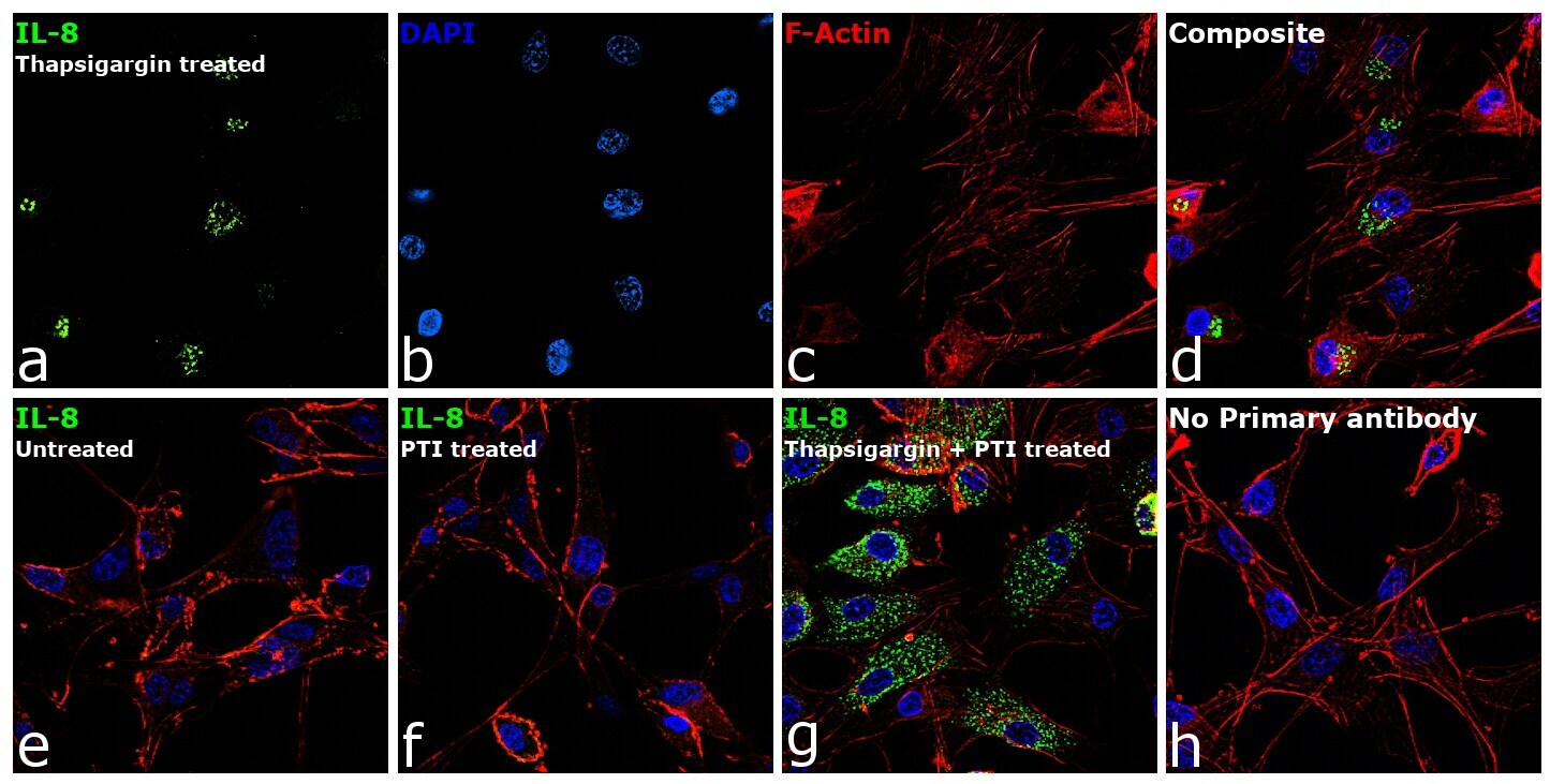
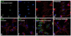
- Immunofluorescence analysis of IL-8 (1-77) (CXCL8) was performed using 70% confluent log phase U-87 MG cells treated with 1uM of Thapsigargin for 24 hours. The cells were fixed with 4% paraformaldehyde for 10 minutes, permeabilized with 0.1% Triton™ X-100 for 15 minutes, and blocked with 2% BSA for 45 minutes at room temperature. The cells were labeled with IL-8 (1-77) (CXCL8) Monoclonal Antibody (N11), eBioscience™ (Product # 14-7189-82) at 5 µg/mL in 0.1% BSA, incubated at 4 degree celsius overnight and then labeled with Goat anti-Mouse IgG (H+L) Highly Cross-Adsorbed Secondary Antibody, Alexa Fluor Plus 488 (Product # A32723), (1:2000), for 45 minutes at room temperature (Panel a: Green). Nuclei (Panel b:Blue) were stained with ProLong™ Diamond Antifade Mountant with DAPI (Product # P36962). F-actin (Panel c: Red) was stained with Rhodamine Phalloidin (Product # R415, 1:300). Panel d represents the merged image showing cytoplasmic(Golgi complex like pattern) localization. Panel e represents untreated cells . Panel f represents cells treated with PTI . Panel g represents cells treated with Thapsigargin and PTI. Panel h represents control cells with no primary antibody to assess background. The images were captured at 60X magnification.