Explore
 Validate
 Learn
Learn Western blot
Antibody data
- Antibody Data
- Antigen structure
- References [0]
- Comments [0]
- Validations
- Western blot [2]
- Immunohistochemistry [1]
- Flow cytometry [1]
Submit
Validation data
Reference
Comment
Report error
- Product number
- AGR-043-200UL - Provider product page

- Provider
- Invitrogen Antibodies
- Product name
- GPR65 (TDAG8) (extracellular) Polyclonal Antibody
- Antibody type
- Polyclonal
- Antigen
- Other
- Reactivity
- Human, Mouse, Rat
- Host
- Rabbit
- Isotype
- IgG
- Vial size
- 200 µL
- Concentration
- 0.8 mg/mL
- Storage
- -20° C, Avoid Freeze/Thaw Cycles
No comments: Submit comment
Supportive validation
- Submitted by
- Invitrogen Antibodies (provider)
- Main image

- Experimental details
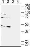
- Western blot analysis of rat (lanes 1 and 3) and mouse (lanes 2 and 4)brain membranes: - 1,2. Anti-GPR65 (TDAG8) (extracellular) Antibody (#AGR-043), (1:200).3,4. Anti-GPR65 (TDAG8) (extracellular) Antibody , preincubated with GPR65/TDAG8 (extracellular) Blocking Peptide (#BLP-GR043).
- Submitted by
- Invitrogen Antibodies (provider)
- Main image
- Experimental details
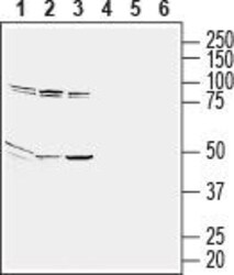
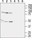
- Western blot analysis of human Jurkat T-cell leukemia (lanes 1 and 4), human THP-1 monocytic leukemia (lanes 2 and 5) and human HL-60 promyelocytic leukemia (lanes 3 and 6) cell line lysates: - 1-3. Anti-GPR65 (TDAG8) (extracellular) Antibody (#AGR-043), (1:200).4-6. Anti-GPR65 (TDAG8) (extracellular) Antibody , preincubated with GPR65/TDAG8 (extracellular) Blocking Peptide (#BLP-GR043).
Supportive validation
- Submitted by
- Invitrogen Antibodies (provider)
- Main image
- Experimental details
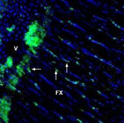
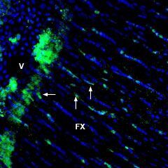
- Expression of GPR65 in rat fornix - Immunohistochemical staining of immersion-fixed, free floating rat frozen brain sections using Anti-GPR65 (TDAG8) (extracellular) Antibody (#AGR-043), (1:1200), followed by goat- Anti-rabbit-AlexaFluor-488. GPR65 staining (green) appears in glial processes (vertical arrows) and in the side of fornix (FX) facing the ventricle (V, horizontal arrow). Cell nuclei are stained with DAPI (blue).
Supportive validation
- Submitted by
- Invitrogen Antibodies (provider)
- Main image
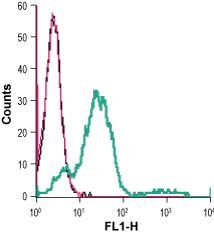
- Experimental details
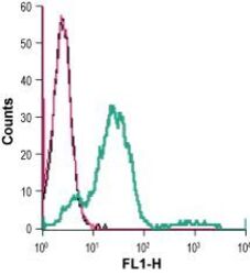
- Cell surface detection of GPR65 in live intact human Jurkat T-cell leukemia cells: - (black line) cells. (red) Cells + goat- Anti-rabbit-FITC. (green) Cells + Anti-GPR65 (TDAG8) (extracellular) Antibody (#AGR-043), 5 µg + goat- Anti-rabbit-FITC.