Explore
 Validate
 Learn
Learn Western blot
Antibody data
- Antibody Data
- Antigen structure
- References [0]
- Comments [0]
- Validations
- Western blot [1]
- Immunocytochemistry [1]
Submit
Validation data
Reference
Comment
Report error
- Product number
- ABIN2508192 - Provider product page

- Provider
- antibodies-online
- Product name
- anti-GCP-2 antibody
- Antibody type
- Polyclonal
- Antigen
- Other
- Description
- Produced from sera of rabbits pre-immunized with highly pure (>98%) recombinant hGCP-2. Anti-Human GCP-2 specific antibody was purified by affinity chromatography employing immobilized hGCP-2 matrix.
- Reactivity
- Human
- Host
- Rabbit
- Vial size
- 100 μg
- Storage
- -20°C
No comments: Submit comment
Supportive validation
- Submitted by
- antibodies-online (provider)
- Main image
- Experimental details
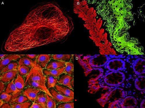
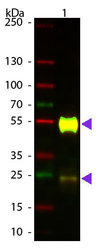
- Western Blot of ATTO 594 conjugated Goat anti-Mouse IgG (Pre-Adsorbed) secondary antibody. Lane 1: Mouse IgG. Lane 2: none. Load: 50 ng per lane. Primary antibody: none. Secondary antibody: ATTO 594 goat secondary antibody at 1:1,000 for 60 min at RT. Block: MB-070 for 30 min at RT. Predicted/Observed size: 25 & 55 kDa, 25 & 55 kDa for Mouse IgG. Other band(s): none. ATTO ® dyes can be used for multicolor immunofluorescent detection with low background and high signal. Examples shown are: A. Tubulin in PtK2- male Rat Kangaroo Kidney Epithelial Cells was detected using ATTO 532 labeled secondary antibody. B. Muscle alpha-actin was stained with a mouse primary antibody and ATTO 488 anti-mouse IgG (green) while Cytokeratin was stained with polyclonal rabbit anti-cytokeratin and ATTO 647N anti-rabbit IgG (red). C. HUVEC (Human umbilical vein endothelial cells were stained with anti- Vimentin-ATTO 532 (green), anti-E-Cadherin-ATTO 655 (red) and DAPI (blue). D. Rat colon sections were stained with Anti-Aquaporin 3-ATTO 594 antibody. Hoechst 33342 (blue) is used as counterstain. Images provided courtesy of Dr. Jörg Reichwein, ATTO-TEC GmbH

Supportive validation
- Submitted by
- antibodies-online (provider)
- Main image

- Experimental details
- Image(s): Immunofluorescence