Explore
 Validate
 Learn
Learn700656
antibody from Invitrogen Antibodies
Targeting: CXCL5
ENA-78, SCYB5
 Western blot
 ELISA Immunocytochemistry Immunoprecipitation Immunohistochemistry Flow cytometry Other assay
Antibody data
- Antibody Data
- Antigen structure
- References [0]
- Comments [0]
- Validations
- ELISA [1]
- Immunocytochemistry [1]
- Immunoprecipitation [1]
- Immunohistochemistry [1]
- Other assay [1]
Submit
Validation data
Reference
Comment
Report error
- Product number
- 700656 - Provider product page

- Provider
- Invitrogen Antibodies
- Product name
- CXCL5 Recombinant Rabbit Monoclonal Antibody (9H45L5)
- Antibody type
- Monoclonal
- Antigen
- Other
- Description
- Intact IgG appears on a non-reducing gel as ~150 kDa band and upon reduction generating a ~25 kDa light chain band and a ~50 kDa heavy chain. Recombinant rabbit monoclonal antibodies are produced using in vitro expression systems. The expression systems are developed by cloning in the specific antibody DNA sequences from immunoreactive rabbits. Then, individual clones are screened to select the best candidates for production. The advantages of using recombinant rabbit monoclonal antibodies include: better specificity and sensitivity, lot-to-lot consistency, animal origin-free formulations, and broader immunoreactivity to diverse targets due to larger rabbit immune repertoire.
- Reactivity
- Human
- Host
- Rabbit
- Isotype
- IgG
- Antibody clone number
- 9H45L5
- Vial size
- 100 μg
- Concentration
- 0.5 mg/mL
- Storage
- Store at 4°C short term. For long term storage, store at -20°C, avoiding freeze/thaw cycles.
No comments: Submit comment
Supportive validation
- Submitted by
- Invitrogen Antibodies (provider)
- Main image

- Experimental details
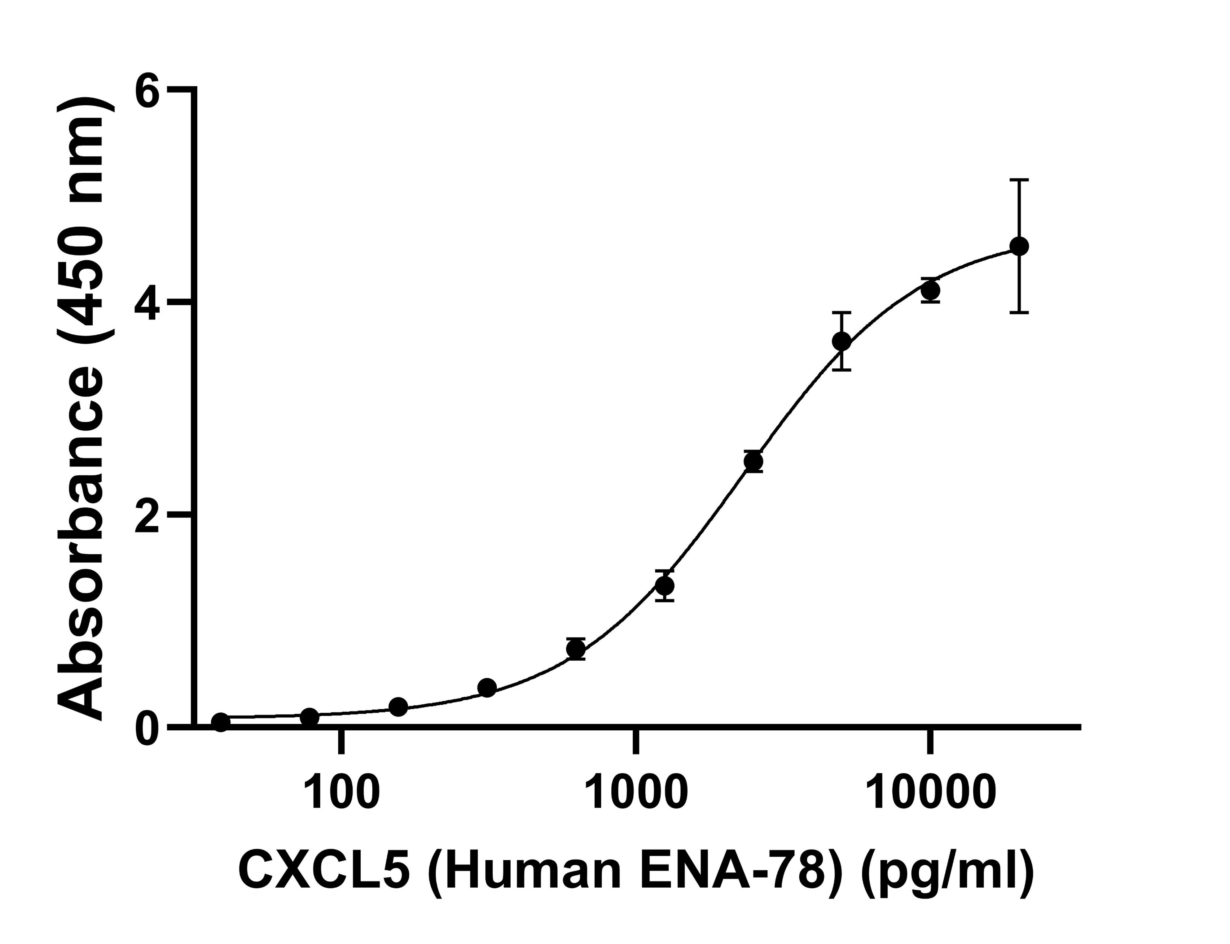
- CXCL5 Recombinant Rabbit Monoclonal Antibody (9H45L5) (Product # 700656) was analyzed by Indirect ELISA. The assay was performed by coating Human ENA-78 (CXCL5) (5-78 aa) Recombinant Protein, PeproTech® (Product # 300-22-20UG) at concentrations of 3.9, 7.8, 15.6, 31.3, 62.5, 125, 250, 500, 1000 and 2000 pg/well. These antigens were incubated with CXCL5 Recombinant Rabbit Monoclonal Antibody (9H45L5) (Product # 700656) (100 ng/well) and detected using Goat anti-Rabbit IgG (H+L) Superclonal™ Recombinant Secondary Antibody, HRP (Product # A27036, 1:5,000). The plate was developed using TMB stabilized chromogen solution (Product # SB02). The plate was read at 450 nm with Thermo Scientific™ Varioskan™ LUX multimode microplate reader (Product # VLBLATD2).
Supportive validation
- Submitted by
- Invitrogen Antibodies (provider)
- Main image
- Experimental details
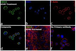
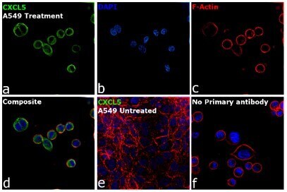
- Immunofluorescence analysis of CXCL5 was performed using 70% confluent log phase A549 cells treated with serum starvation (16 hours), PMA (10 nM for 24 hours), TNFa (10 ng/mL for 24 hours) and BFA (300 ng/mL for last 3 hours). The cells were fixed with 4% paraformaldehyde for 10 minutes, permeabilized with 0.1% Triton™ X-100 for 15 minutes, and blocked with 2% BSA for 1 hour at room temperature. The cells were labeled with CXCL5 Recombinant Rabbit Monoclonal Antibody (9H45L5) (Product # 700656) at 1 µg/mL in 0.1% BSA, incubated at 4 degree celsius overnight and then labeled with Donkey anti-Rabbit IgG (H+L) Highly Cross-Adsorbed Secondary Antibody, Alexa Fluor™ Plus 488 (Product # A32790, 1:2,000), for 45 minutes at room temperature (Panel a: Green). Nuclei (Panel b:Blue) were stained with ProLong™ Diamond Antifade Mountant with DAPI (Product # P36962). F-actin (Panel c: Red) was stained with Rhodamine Phalloidin (Product # R415, 1:300). Panel d represents the merged image showing cytoplasmic localization. Panel e represents untreated A549 cells with no signal intensity as compared to the treated cells. Panel f represents control cells with no primary antibody to assess the background. The images were captured at 40X magnification.
Supportive validation
- Submitted by
- Invitrogen Antibodies (provider)
- Main image

- Experimental details
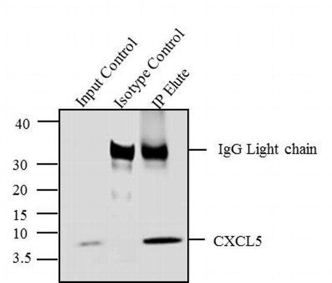
- CXCL5 was immunoprecipitated using 5 µg of the CXCL5 Recombinant Rabbit Monoclonal Antibody (Product # 700656) from lysate of A549 cells treated with PMA (200 nM/20 minutes) (Lane 3) using the Dynabeads® Protein A Immunoprecipitation Kit (Product # 10006D). Normal Rabbit IgG was used as a Isotype control (Lane 2). 10 % input represents the cell extract used for immunoprecipitation (Lane 1). Western blot analysis was performed using CXCL5 Recombinant Rabbit Monoclonal Antibody (Product # 700656) and Goat anti-Rabbit IgG (Heavy Chain) Superclonal™ Secondary Antibody, HRP conjugate (Product # A27036, 0.4 µg/mL, 1:2500 dilution). Chemiluminescent detection was performed using Pierce™ ECL Western blotting Substrate (Product # 32106).

Supportive validation
- Submitted by
- Invitrogen Antibodies (provider)
- Main image
- Experimental details
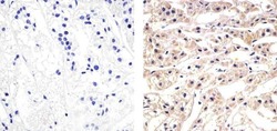
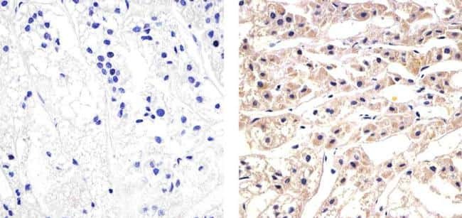
- Immunohistochemistry analysis of CXCL5 showing staining in the cytoplasm of paraffin-embedded human adrenal gland (right) compared to a negative control without primary antibody (left). To expose target proteins, antigen retrieval was performed using 10mM sodium citrate (pH 6.0), microwaved for 8-15 min. Following antigen retrieval, tissues were blocked in 3% H2O2-methanol for 15 min at room temperature, washed with ddH2O and PBS, and then probed with a CXCL5 Recombinant Rabbit Monoclonal Antibody (Product # 700656) diluted in 3% BSA-PBS at a dilution of 1:20 overnight at 4°C in a humidified chamber. Tissues were washed extensively in PBST and detection was performed using an HRP-conjugated secondary antibody followed by colorimetric detection using a DAB kit. Tissues were counterstained with hematoxylin and dehydrated with ethanol and xylene to prep for mounting.
Supportive validation
- Submitted by
- Invitrogen Antibodies (provider)
- Main image

- Experimental details
- CXCL5 was immunoprecipitated using 5 æg of the CXCL5 Recombinant Rabbit Monoclonal Antibody (Product # 700656) from lysate of A549 cells treated with PMA (200 nM/20 minutes) (Lane 3) using the Dynabeads® Protein A Immunoprecipitation Kit (Product # 10006D). Normal Rabbit IgG was used as a Isotype control (Lane 2). 10 % input represents the cell extract used for immunoprecipitation (Lane 1). Western blot analysis was performed using CXCL5 Recombinant Rabbit Monoclonal Antibody (Product # 700656) and Goat anti-Rabbit IgG (H+L) Superclonal™ Secondary Antibody, HRP conjugate (Product # A27036, 0.4 æg/mL, 1:2500 dilution). Chemiluminescent detection was performed using Pierce™ ECL Western blotting Substrate (Product # 32106).