 Explore
 Validate
 Learn
Learn Western blot
 Immunocytochemistry
Antibody data
- Antibody Data
- Antigen structure
- References [3]
- Comments [0]
- Validations
- Immunocytochemistry [1]
- Immunohistochemistry [1]
Submit
Validation data
Reference
Comment
Report error
- Product number
- HPA008001 - Provider product page

- Provider
- Atlas Antibodies
- Proper citation
- Atlas Antibodies Cat#HPA008001, RRID:AB_1853292
- Product name
- Anti-LRPAP1
- Antibody type
- Polyclonal
- Description
- Polyclonal Antibody against Human LRPAP1, Gene description: low density lipoprotein receptor-related protein associated protein 1, Alternative Gene Names: A2MRAP, HBP44, Validated applications: ICC, IHC, WB, Uniprot ID: P30533, Storage: Store at +4°C for short term storage. Long time storage is recommended at -20°C.
- Reactivity
- Human, Mouse, Rat
- Host
- Rabbit
- Conjugate
- Unconjugated
- Isotype
- IgG
- Vial size
- 100 µl
- Concentration
- 0.1 mg/ml
- Storage
- Store at +4°C for short term storage. Long time storage is recommended at -20°C.
- Handling
- The antibody solution should be gently mixed before use.
Submitted references Secreted LRPAP1 binds and triggers IFNAR1 degradation to facilitate virus evasion from cellular innate immunity
Systematic validation of antibody binding and protein subcellular localization using siRNA and confocal microscopy
Tissue profiling of the mammalian central nervous system using human antibody-based proteomics.
Li H, Wang X, Wang Y, Li Y, Chen Y, Wong Y, He J, He M
Signal Transduction and Targeted Therapy 2023;8(1)
Signal Transduction and Targeted Therapy 2023;8(1)
Systematic validation of antibody binding and protein subcellular localization using siRNA and confocal microscopy
Stadler C, Hjelmare M, Neumann B, Jonasson K, Pepperkok R, Uhlén M, Lundberg E
Journal of Proteomics 2012;75(7):2236-2251
Journal of Proteomics 2012;75(7):2236-2251
Tissue profiling of the mammalian central nervous system using human antibody-based proteomics.
Mulder J, Björling E, Jonasson K, Wernérus H, Hober S, Hökfelt T, Uhlén M
Molecular & cellular proteomics : MCP 2009 Jul;8(7):1612-22
Molecular & cellular proteomics : MCP 2009 Jul;8(7):1612-22
No comments: Submit comment
Supportive validation
- Submitted by
- Atlas Antibodies (provider)
- Main image
- Experimental details
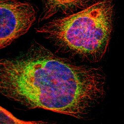
- Immunofluorescent staining of human cell line A-431 shows localization to endoplasmic reticulum.
- Sample type
- Human

Supportive validation
- Submitted by
- Atlas Antibodies (provider)
- Enhanced method
- Orthogonal validation
- Main image
- Experimental details
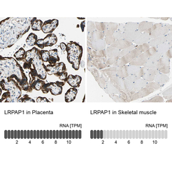
- Immunohistochemistry analysis in human placenta and skeletal muscle tissues using HPA008001 antibody. Corresponding LRPAP1 RNA-seq data are presented for the same tissues.
- Sample type
- Human
- Protocol
- Protocol
