 Explore
 Validate
 Learn
Learn Western blot
Antibody data
- Antibody Data
- Antigen structure
- References [0]
- Comments [0]
- Validations
- Western blot [5]
Submit
Validation data
Reference
Comment
Report error
- Product number
- MA1-26155 - Provider product page

- Provider
- Invitrogen Antibodies
- Product name
- SCD Monoclonal Antibody (CD.E10)
- Antibody type
- Monoclonal
- Antigen
- Recombinant full-length protein
- Description
- Recommended positive controls: 293 cells.
- Antibody clone number
- CD.E10
- Concentration
- 1 mg/mL
No comments: Submit comment
Supportive validation
- Submitted by
- Invitrogen Antibodies (provider)
- Main image

- Experimental details
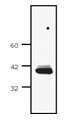
- Western Blot analysis of 293T cell lysate using SCD Monoclonal Antibody (CD. E10) (Product # MA1-26155).
- Submitted by
- Invitrogen Antibodies (provider)
- Main image
- Experimental details
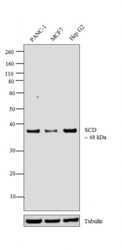
- Western blot analysis was performed on whole cell extracts (30 µg lysate) of PANC-1 (Lane 1), MCF7 (Lane 2) and Hep G2 (Lane 3). The blot was probed with Anti-SCD Monoclonal Antibody (CD.E10) (Product # MA1-26155, 1:1000 dilution) and detected by chemiluminescence using Goat anti-Mouse IgG (H+L) Superclonal™ Secondary Antibody, HRP conjugate (Product # A28177, 0.25µg/ml, 1:4000 dilution). A 48 kDa band corresponding to SCD was observed across the cell lines tested.

- Submitted by
- Invitrogen Antibodies (provider)
- Main image

- Experimental details
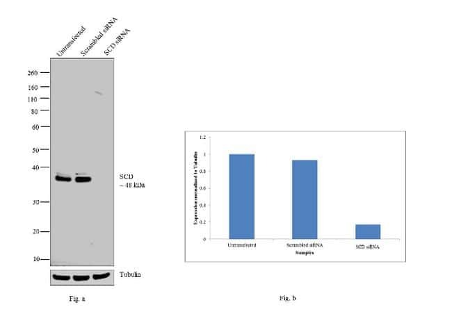
- Knockdown of SCD was achieved by transfecting MCF7 cells with SCD specific siRNAs (Silencer® select Product # s12505). Western blot analysis (Fig. a) was performed using whole cell extracts from the SCD knockdown cells (lane 3), non-specific scrambled siRNA transfected cells (lane 2) and untransfected cells (lane 1). The blot was probed with SCD Monoclonal Antibody (CD.E10) (Product # MA1-26155, 1:1000 dilution) and Goat anti-Mouse IgG (H+L) Superclonal™ Secondary Antibody, HRP conjugate (Product # A28177, 0.25µg/ml, 1:4000 dilution). Densitometric analysis of this western blot is shown in histogram (Fig. b). Decrease in signal upon siRNA mediated knock down confirms that antibody is specific to SCD.
- Submitted by
- Invitrogen Antibodies (provider)
- Main image

- Experimental details
- Western Blot analysis of 293T cell lysate using SCD Monoclonal Antibody (CD. E10) (Product # MA1-26155).

- Submitted by
- Invitrogen Antibodies (provider)
- Main image
- Experimental details
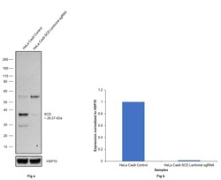
- CRISPR-Cas9 mediated genome editing ofSCD (as confirmed by next generation sequencing) was achieved by using LentiArray™ Lentiviral sgRNA (Product # A32042, Assay ID CRISPR730414_LV) and LentiArray Cas9 Lentivirus (Product # A32064). Fig (a) Western blot analysis of SCD was performed by loading 30 µg of HeLa Cas9 (Lane 1) and HeLa Cas9 cells transduced with SCD Lentiviral sgRNA (Lane 2) whole cell extracts. The samples were electrophoresed using NuPAGE™ Novex™ 4-12% Bis-Tris Protein Gel (Product # NP0322BOX). Resolved proteins were then transferred onto a nitrocellulose membrane (Product # IB23001) by iBlot® 2 Dry Blotting System (Product # IB21001). The blot was probed with Anti-SCD Monoclonal Antibody (CD.E10) (Product # MA1-26155) using 1:1000 dilution and Goat anti-Mouse IgG (H+L) Superclonal™ Recombinant Secondary Antibody, HRP (Product # A28177 1:10000 dilution).Chemiluminescent detection was performed using SuperSignal™ West Dura Extended Duration Substrate (Product # 34076). A reduced signal in sgRNA transduced cells using the LentiArray™ CRISPR product line confirms that antibody is specific toSCD (Fig (b)).
