Explore
 Validate
 Learn
Learn Western blot
Antibody data
- Antibody Data
- Antigen structure
- References [0]
- Comments [0]
- Validations
- Western blot [3]
- Immunohistochemistry [1]
Submit
Validation data
Reference
Comment
Report error
- Product number
- PA5-19239 - Provider product page

- Provider
- Invitrogen Antibodies
- Product name
- SNX1 Polyclonal Antibody
- Antibody type
- Polyclonal
- Antigen
- Synthetic peptide
- Description
- This antibody is predicted to react with bovine, canine, mouse and rat based on sequence homology. This antibody is tested in Peptide ELISA: antibody detection limit dilution 64,000.
- Reactivity
- Human
- Host
- Goat
- Isotype
- IgG
- Vial size
- 100 µg
- Concentration
- 0.5 mg/mL
- Storage
- -20° C, Avoid Freeze/Thaw Cycles
No comments: Submit comment
Supportive validation
- Submitted by
- Invitrogen Antibodies (provider)
- Main image

- Experimental details
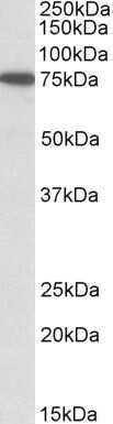
- Western Blot staining of HeLa cell lysate using Product # PA5-19239 at a concentration of 1.0 µg/mL, the primary antibody incubation was 1 hour and the detection method was chemiluminescence.
- Submitted by
- Invitrogen Antibodies (provider)
- Main image

- Experimental details
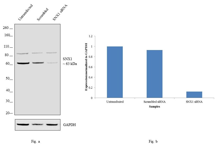
- KD of SNX1 was achieved by transfecting HeLa with SNX1 specific siRNAs (Silencer® select Product # s13257, s13526). Western blot analysis (Fig. a) was performed using membrane enriched cell extracts from the SNX1 KD cells (Lane 3), non-specific scrambled siRNA transfected cells (Lane 2) and untransfected cells (Lane 1). The blot was probed with SNX1 Polyclonal Antibody (Product # PA5-19239, 1 µg/mL) and Rabbit anti-Goat IgG (H+L) Superclonal™ Secondary Antibody, HRP conjugate (Product # A27014, 0.25µg/mL, 1:4000 dilution). Densitometric analysis of this western blot is shown in histogram (Fig. b). Decrease in signal upon siRNA mediated knock down confirms that antibody is specific to SNX1.
- Submitted by
- Invitrogen Antibodies (provider)
- Main image
- Experimental details
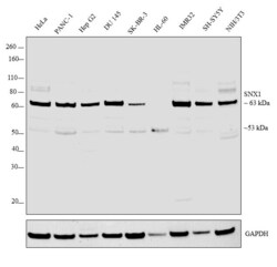
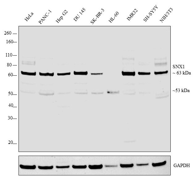
- Western blot was performed using Anti-SNX1 Goat Polyclonal Antibody (Product # PA5-19239) and a 63 kDa band corresponding to SNX1 was observed across cell lines tested. Membrane enriched cell extracts (30 µg lysate) of HeLa (Lane 1), PANC-1 (Lane 2), Hep G2 (Lane 3), DU 145 (Lane 4), SK-BR-3 (Lane 5), HL-60 (Lane 6), IMR32 (Lane 7), SH-SY5Y (Lane 8) and NIH/3T3 (Lane 9) were electrophoresed using Novex® NuPAGE® 4-12 % Bis-Tris gel (Product # NP0321BOX). Resolved proteins were then transferred onto a nitrocellulose membrane (Product # IB23001) by iBlot® 2 Dry Blotting System (Product # IB21001). The blot was probed with the primary antibody (1 µg/mL) and detected by chemiluminescence Rabbit Anti-Goat IgG Secondary Antibody, HRP conjugate (Product # A27014, 1:4000 dilution) using the iBright FL 1000 (Product # A32752). Chemiluminescent detection was performed using Novex® ECL Chemiluminescent Substrate Reagent Kit (Product # WP20005).
Supportive validation
- Submitted by
- Invitrogen Antibodies (provider)
- Main image

- Experimental details
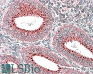
- Immunohistochemical analysis of SNX1 in paraffin embedded human uterus using a SNX1 polyclonal antibody (Product #PA5-19239) at a concentration of 3.8 µg/mL. Steamed antigen retrieval was performed with pH 6 buffered citrate. Samples were then stained with alkaline phosphatase.