Explore
 Validate
 Learn
Learn Western blot
 Immunocytochemistry
Antibody data
- Antibody Data
- Antigen structure
- References [1]
- Comments [0]
- Validations
- Immunocytochemistry [2]
- Immunohistochemistry [1]
- Other assay [3]
Submit
Validation data
Reference
Comment
Report error
- Product number
- PA5-34745 - Provider product page

- Provider
- Invitrogen Antibodies
- Product name
- LIMCH1 Polyclonal Antibody
- Antibody type
- Polyclonal
- Antigen
- Recombinant full-length protein
- Description
- Recommended positive controls: H1299, HeLaS3, mouse brain. Predicted reactivity: Mouse (80%), Bovine (86%). IHC notes, Requires antigen retrieval using heat mediated 10mM Citrate buffer (pH6.0) or Tris-EDTA buffer (pH8.0) Store product as a concentrated solution. Centrifuge briefly prior to opening the vial.
- Reactivity
- Human, Mouse
- Host
- Rabbit
- Isotype
- IgG
- Vial size
- 100 μL
- Concentration
- 1.53 mg/mL
- Storage
- Store at 4°C short term. For long term storage, store at -20°C, avoiding freeze/thaw cycles.
Submitted references Circular RNA circLDB2 functions as a competing endogenous RNA to suppress development and promote cisplatin sensitivity in non-squamous non-small cell lung cancer.
Wang Y, Li L, Zhang W, Zhang G
Thoracic cancer 2021 Jul;12(13):1959-1972
Thoracic cancer 2021 Jul;12(13):1959-1972
No comments: Submit comment
Supportive validation
- Submitted by
- Invitrogen Antibodies (provider)
- Main image

- Experimental details
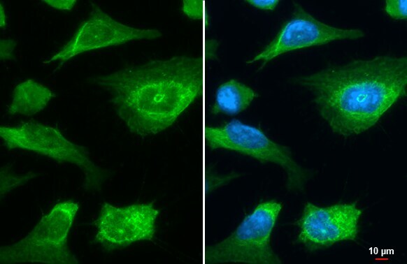
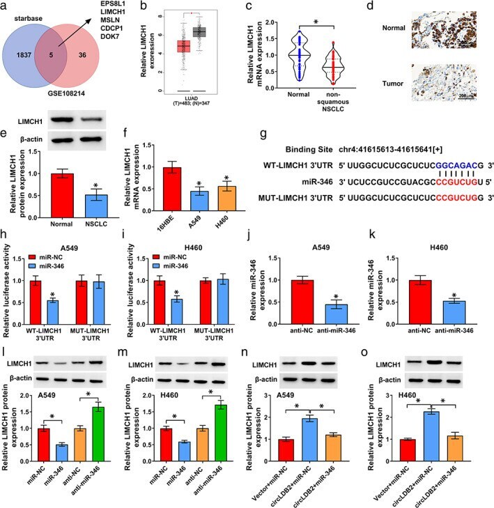
- LIMCH1 Polyclonal Antibody detects LIMCH1 protein at stress fiber by immunofluorescent analysis. Sample: HeLa cells were fixed in ice-cold MeOH for 5 min. Green: LIMCH1 stained by LIMCH1 Polyclonal Antibody (Product # PA5-34745) diluted at 1:500.
- Submitted by
- Invitrogen Antibodies (provider)
- Main image
- Experimental details
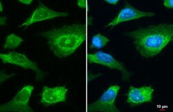
- LIMCH1 Polyclonal Antibody detects LIMCH1 protein at stress fiber by immunofluorescent analysis. Sample: HeLa cells were fixed in ice-cold MeOH for 5 min. Green: LIMCH1 stained by LIMCH1 Polyclonal Antibody (Product # PA5-34745) diluted at 1:500.

Supportive validation
- Submitted by
- Invitrogen Antibodies (provider)
- Main image

- Experimental details
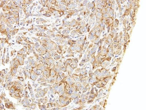
- Immunohistochemical analysis of paraffin-embedded A549 xenograft, using LIMCH1 (Product # PA5-34745) antibody at 1:500 dilution. Antigen Retrieval: Citrate buffer, pH 6.0, 15 min.
Supportive validation
- Submitted by
- Invitrogen Antibodies (provider)
- Main image
- Experimental details
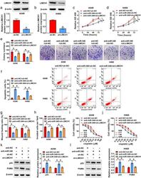
- FIGURE 7 LIMCH1 was a functional target of miR-346. (a) and (b) Western blot analysis of LIMCH1 protein level in cells transfected with sh-NC or sh-LIMCH1. A549 and H460 cells were transfected with anti-NC + sh-NC, anti-miR-346 + sh-NC or anti-miR-346 + sh-LIMCH1. (c) and (d) CCK-8 assay of proliferation of transfected cells. (e) Representative images depicting a colony formation assay. (f) Representative images depicting a cell apoptosis assay and cell apoptosis by flow cytometry. (g) Wound-healing assay of cell migration. (h) Transwell assay of cell invasion. (i) and (j) CCK-8 assay for viability of transfected cells under cisplatin exposure. (k) and (l) Western blot showing the levels of p53 and PUMA in transfected cells. * p < 0.05
- Submitted by
- Invitrogen Antibodies (provider)
- Main image

- Experimental details
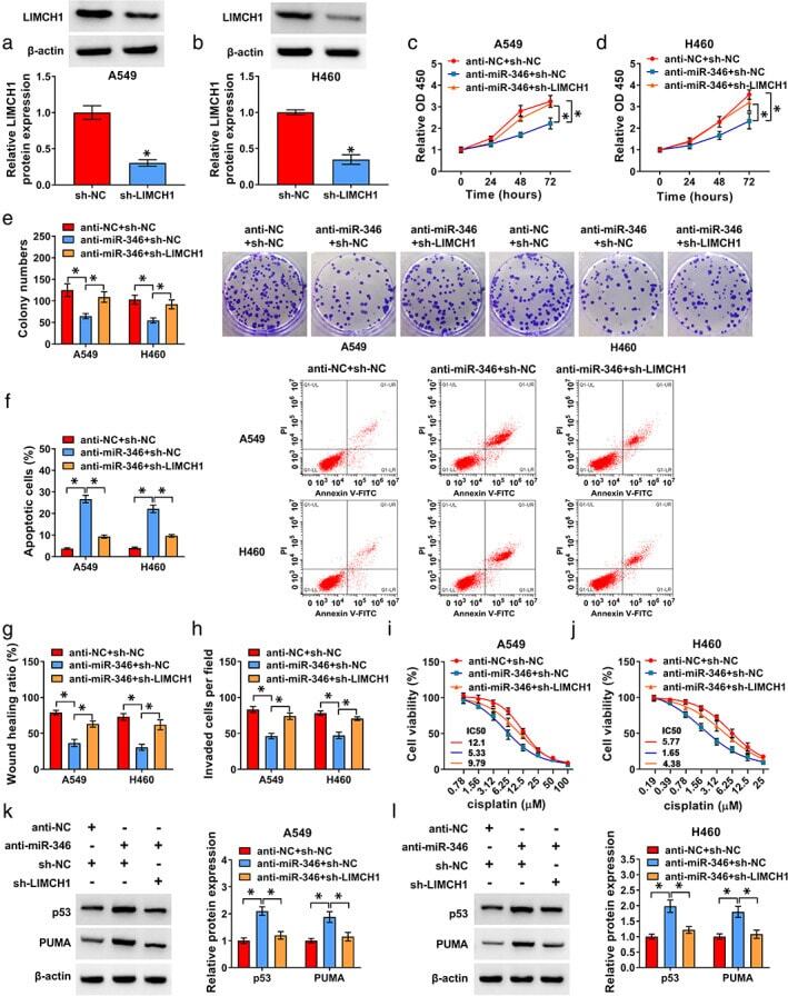
- FIGURE 6 CircLDB2 modulated LIMCH1 expression by acting as a ceRNA for miR-346. (a) Venn diagram showing the putative targets of miR-346 identified by starbase software and GEO database (GSE108214). (b) the expression of LIMCH1 in LUAD and LUSC tissues compared with the controls analyzed by the GEPIA database. (c) qRT-PCR analysis of LIMCH1 mRNA in 53 primary tumors and adjacent noncancerous lung tissues from the same patients with non-squamous NSCLC. (d) Representative images depicting an immunohistochemistry assay of LIMCH1 protein expression in tissue samples. (e) Representative images depicting a western blot assay and western blot showing LIMCH1 protein in three pairs of tumor samples and adjacent noncancerous lung tissues. (f) qRT-PCR analysis showing the expression of LIMCH1 mRNA in 16HBE, A549, and H460 cells. (g) Sequence of miR-346, the miR-346 binding sites within LIMCH1 3'UTR and the mutant of the target region. (h) and (i) dual-luciferase reporter assays in A549 and H460 cells with LIMCH1 3'UTR wild-type (WT-LIMCH1 3'UTR) or mutant (MUT-LIMCH1 3'UTR) luciferase reporter constructs. (j) and (k) qRT-PCR analysis of miR-346 expression in anti-NC- or anti-miR-346-transfected A549 and H460 cells. (l) and (m) Western blot showing the expression of LIMCH1 protein in cells transfected with miR-NC mimic, miR-346 mimic, anti-NC, or anti-miR-346. (n) and (o) LIMCH1 protein level by western blot in vector-transduced or circLDB2-transduced cells transfected with miR-NC mimic
- Submitted by
- Invitrogen Antibodies (provider)
- Main image
- Experimental details
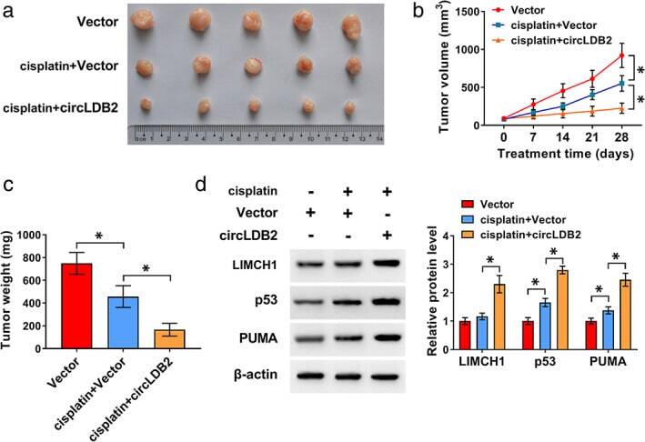
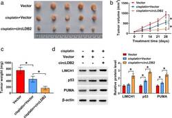
- FIGURE 8 Overexpression of circLDB2 promoted the antitumor effect of cisplatin in vivo. The xenograft tumors were generated by subcutaneous injection of vector-transduced or circLDB2-transduced A549 cells (2 x 10 6 per mouse, n = 5 per group), and cisplatin treatment was performed twice a week by intraperitoneal injection. Mice were terminated at day 28 after cell implantation. (a) Representative images of the xenograft tumors. (b) Growth curves of the excised tumors. (c) Tumor average weight of the xenograft tumors. (d) Western blot showing the levels of LIMCH1, p53, and PUMA in the excised tumors. * p < 0.05