Explore
 Validate
 Learn
Learn Immunocytochemistry
Antibody data
- Antibody Data
- Antigen structure
- References [5]
- Comments [0]
- Validations
- Immunocytochemistry [1]
- Immunohistochemistry [1]
Submit
Validation data
Reference
Comment
Report error
- Product number
- HPA018304 - Provider product page

- Provider
- Atlas Antibodies
- Proper citation
- Atlas Antibodies Cat#HPA018304, RRID:AB_1849350
- Product name
- Anti-G3BP2
- Antibody type
- Polyclonal
- Description
- Polyclonal Antibody against Human G3BP2, Gene description: GTPase activating protein (SH3 domain) binding protein 2, Alternative Gene Names: KIAA0660, Validated applications: IHC, ICC, Uniprot ID: Q9UN86, Storage: Store at +4°C for short term storage. Long time storage is recommended at -20°C.
- Reactivity
- Human
- Host
- Rabbit
- Conjugate
- Unconjugated
- Isotype
- IgG
- Vial size
- 100 µl
- Concentration
- 0.3 mg/ml
- Storage
- Store at +4°C for short term storage. Long time storage is recommended at -20°C.
- Handling
- The antibody solution should be gently mixed before use.
Submitted references Nsp1 proteins of human coronaviruses HCoV-OC43 and SARS-CoV2 inhibit stress granule formation
NAP1L1 and NAP1L4 Binding to Hypervariable Domain of Chikungunya Virus nsP3 Protein Is Bivalent and Requires Phosphorylation
UV damage induces G3BP1-dependent stress granule formation that is not driven by mTOR inhibition-mediated translation arrest
Raloxifene prevents stress granule dissolution, impairs translational control and promotes cell death during hypoxia in glioblastoma cells
Targeting of Rac GTPases blocks the spread of intact human breast cancer
Maier H, Dolliver S, Kleer M, Bui-Marinos M, Ying S, Corcoran J, Khaperskyy D
PLOS Pathogens 2022;18(12):e1011041
PLOS Pathogens 2022;18(12):e1011041
NAP1L1 and NAP1L4 Binding to Hypervariable Domain of Chikungunya Virus nsP3 Protein Is Bivalent and Requires Phosphorylation
Dominguez F, Shiliaev N, Lukash T, Agback P, Palchevska O, Gould J, Meshram C, Prevelige P, Green T, Agback T, Frolova E, Frolov I, López S
Journal of Virology 2021;95(16)
Journal of Virology 2021;95(16)
UV damage induces G3BP1-dependent stress granule formation that is not driven by mTOR inhibition-mediated translation arrest
Ying S, Khaperskyy D
Journal of Cell Science 2020;133(20)
Journal of Cell Science 2020;133(20)
Raloxifene prevents stress granule dissolution, impairs translational control and promotes cell death during hypoxia in glioblastoma cells
Attwood K, Robichaud A, Westhaver L, Castle E, Brandman D, Balgi A, Roberge M, Colp P, Croul S, Kim I, McCormick C, Corcoran J, Weeks A
Cell Death & Disease 2020;11(11)
Cell Death & Disease 2020;11(11)
Targeting of Rac GTPases blocks the spread of intact human breast cancer
Katz E, Sims A, Sproul D, Caldwell H, Dixon J, Meehan R, Harrison D
Oncotarget 2012;3(6):608-619
Oncotarget 2012;3(6):608-619
No comments: Submit comment
Supportive validation
- Submitted by
- Atlas Antibodies (provider)
- Main image
- Experimental details
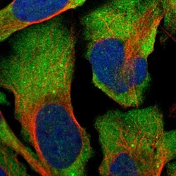
- Immunofluorescent staining of human cell line U-2 OS shows localization to cytosol.
- Sample type
- Human

Supportive validation
- Submitted by
- Atlas Antibodies (provider)
- Enhanced method
- Orthogonal validation
- Main image
- Experimental details
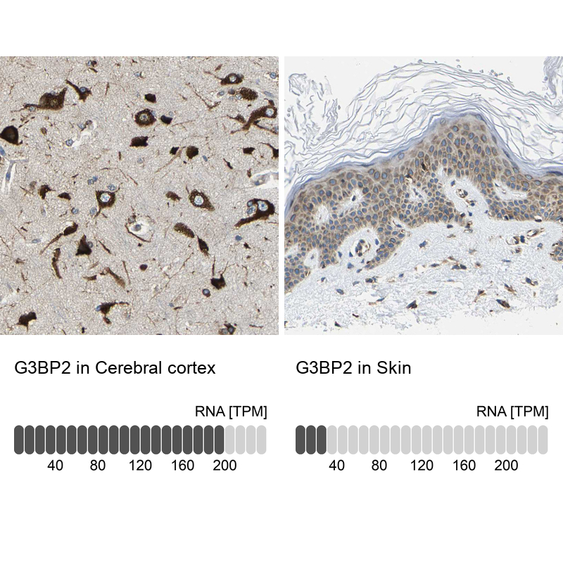
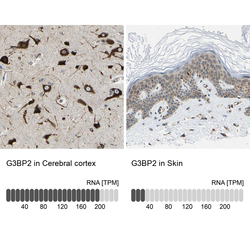
- Immunohistochemistry analysis in human cerebral cortex and skin tissues using HPA018304 antibody. Corresponding G3BP2 RNA-seq data are presented for the same tissues.
- Sample type
- Human
- Protocol
- Protocol