Explore
 Validate
 Learn
Learn Immunocytochemistry
 Immunohistochemistry
Antibody data
- Antibody Data
- Antigen structure
- References [3]
- Comments [0]
- Validations
- Immunocytochemistry [1]
- Immunohistochemistry [1]
Submit
Validation data
Reference
Comment
Report error
- Product number
- HPA018425 - Provider product page

- Provider
- Atlas Antibodies
- Proper citation
- Atlas Antibodies Cat#HPA018425, RRID:AB_1849352
- Product name
- Anti-G3BP2
- Antibody type
- Polyclonal
- Description
- Polyclonal Antibody against Human G3BP2, Gene description: GTPase activating protein (SH3 domain) binding protein 2, Alternative Gene Names: KIAA0660, Validated applications: IHC, Uniprot ID: Q9UN86, Storage: Store at +4°C for short term storage. Long time storage is recommended at -20°C.
- Reactivity
- Human
- Host
- Rabbit
- Conjugate
- Unconjugated
- Isotype
- IgG
- Vial size
- 100 µl
- Concentration
- 0.1 mg/ml
- Storage
- Store at +4°C for short term storage. Long time storage is recommended at -20°C.
- Handling
- The antibody solution should be gently mixed before use.
Submitted references
The miR‐669a‐5p/G3BP/HDAC6/AKAP12 Axis Regulates Primary Cilia Length
Matrix stiffness drives epithelial–mesenchymal transition and tumour metastasis through a TWIST1–G3BP2 mechanotransduction pathway
Kumar A, Tanaka K, Schwartz M
2024
2024
The miR‐669a‐5p/G3BP/HDAC6/AKAP12 Axis Regulates Primary Cilia Length
Wang W, Dai X, Li Y, Li M, Chi Z, Hu X, Wang Z
Advanced Science 2023;11(6)
Advanced Science 2023;11(6)
Matrix stiffness drives epithelial–mesenchymal transition and tumour metastasis through a TWIST1–G3BP2 mechanotransduction pathway
Wei S, Fattet L, Tsai J, Guo Y, Pai V, Majeski H, Chen A, Sah R, Taylor S, Engler A, Yang J
Nature Cell Biology 2015;17(5):678-688
Nature Cell Biology 2015;17(5):678-688
No comments: Submit comment
Enhanced validation
- Submitted by
- 55af80e3e0991
- Enhanced method
- Genetic validation
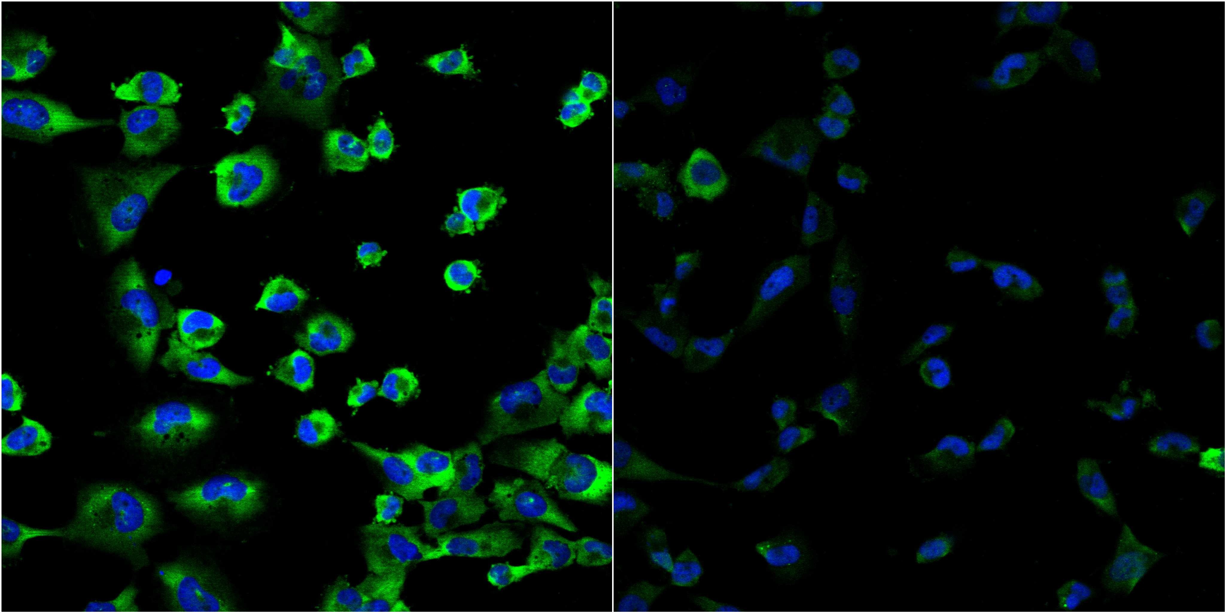
- Main image
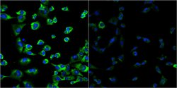
- Experimental details
- Confocal images of immunofluorescently stained human U-2 OS cells.The protein G3BP2 is shown in green and the nucleus in blue. The image to the left show cells transfected with control siRNA and the image to the right show cells where G3BP2 has been downregulated with specific siRNA.
- Sample type
- U-2 OS cells
- Primary Ab dilution
- 1:70
- Secondary Ab
- Secondary Ab
- Secondary Ab dilution
- 1:800
- Knockdown/Genetic Approaches Application
- Immunocytochemistry
Supportive validation
- Submitted by
- Atlas Antibodies (provider)
- Enhanced method
- Orthogonal validation
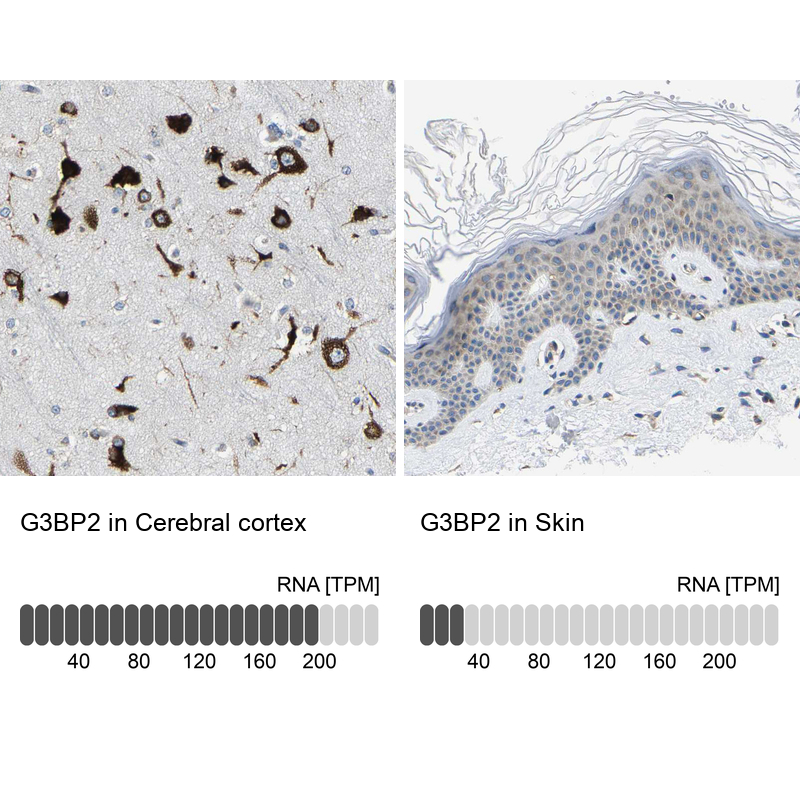
- Main image
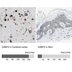
- Experimental details
- Immunohistochemistry analysis in human cerebral cortex and skin tissues using HPA018425 antibody. Corresponding G3BP2 RNA-seq data are presented for the same tissues.
- Sample type
- Human
- Protocol
- Protocol